
Neural Collection
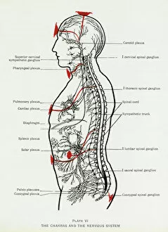
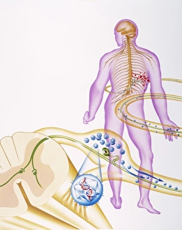
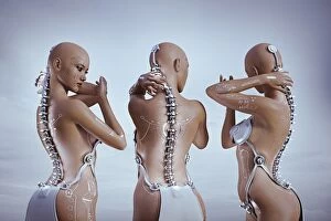
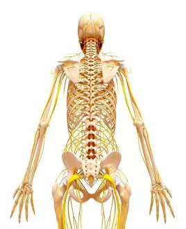
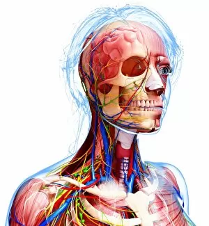
"Unlocking the Mysteries of the Neural Network: Exploring Chakras, Nervous System, and Brain Pathways" Delving into the intricate web of our neural system


All Professionally Made to Order for Quick Shipping
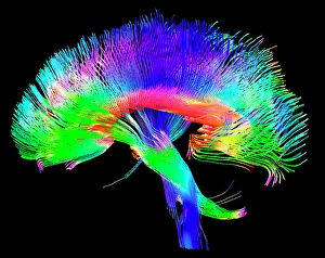
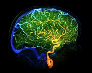
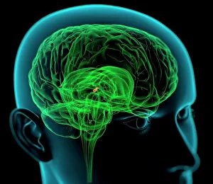
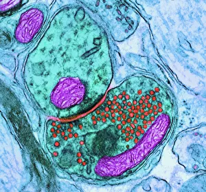
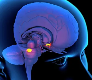
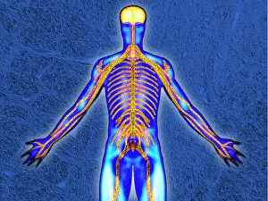
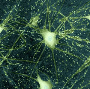
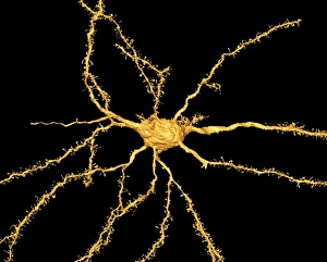
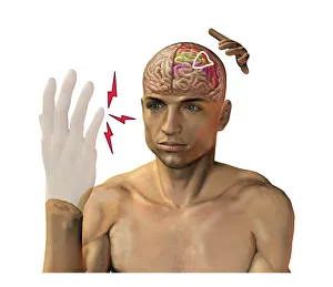
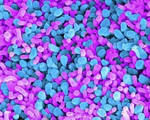
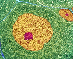
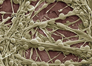
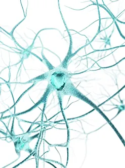
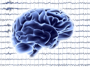
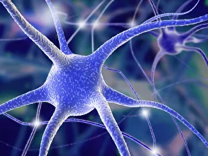
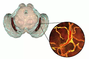
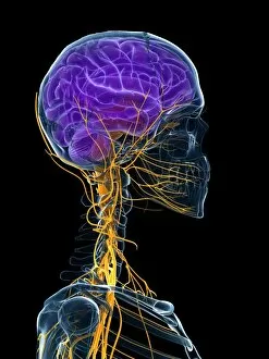
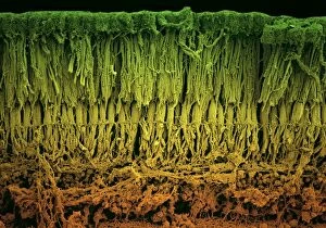
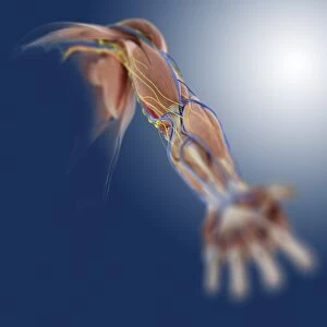
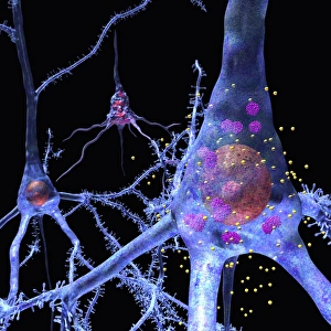
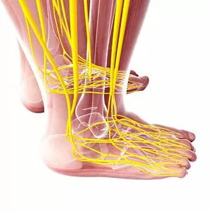
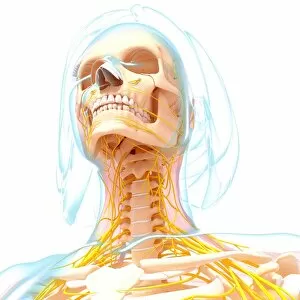
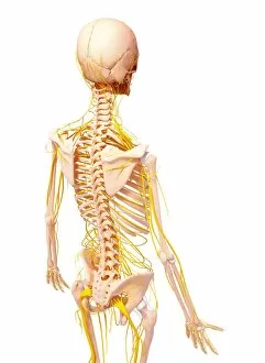
"Unlocking the Mysteries of the Neural Network: Exploring Chakras, Nervous System, and Brain Pathways" Delving into the intricate web of our neural system, we uncover a fascinating realm where chakras intertwine with nerve cells. Like energetic gateways, these chakras facilitate communication between our physical and spiritual selves. Tracing the pathways within our brain, we witness a mesmerizing dance of electrical signals. These brain pathways serve as highways for information exchange, allowing us to perceive the world around us and make sense of it all. Intriguingly, 3D angiograms from 1981 reveal an astonishing view of brain blood vessels. This captivating imagery showcases their intricate network that nourishes every corner of our cognitive powerhouse. The medulla oblongata takes center stage in this artistic representation within the brain. Known as the control center for vital functions like breathing and heart rate regulation, its significance cannot be overstated. Zooming closer into microscopic wonders, we encounter synapse nerve junctions captured through transmission electron microscopy (TEM). These tiny connections are where messages leap across gaps to ensure seamless communication between neurons. Examining nerve cells under scanning electron microscopy (SEM), their complex structures come alive before our eyes. Each cell is a marvel on its own - branching out like trees in a vast forest yet intricately connected to form an awe-inspiring network. Returning to another artistic depiction of the medulla oblongata reinforces its importance within our nervous system's architecture. It serves as a reminder that even amidst scientific exploration lies room for creative interpretation and appreciation. Venturing further into this enigmatic realm reveals more about brain blood vessels' role in supporting optimal cognitive function. Their delicate balance ensures oxygen-rich blood reaches every nook and cranny within this remarkable organ. Light micrographs showcasing motor neurons shed light on how they orchestrate movement throughout our bodies.