
Oncological Collection
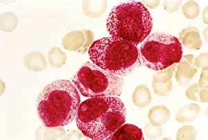
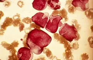
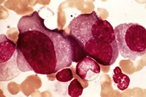
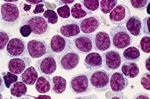
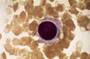
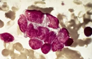
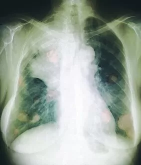
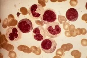
"Unveiling the Hidden Battles: A Glimpse into the World of Oncology" Acute promyelocytic leukaemia, micrograph: Witnessing the intricate battle within


All Professionally Made to Order for Quick Shipping
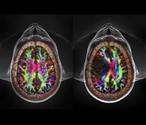
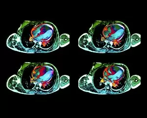
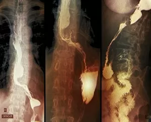
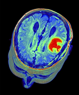
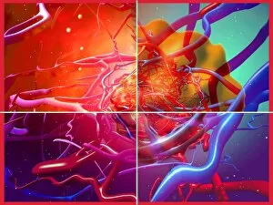
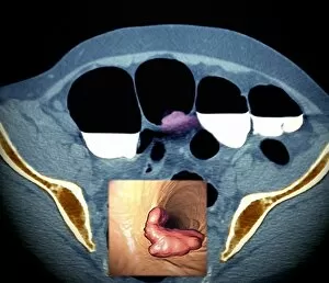
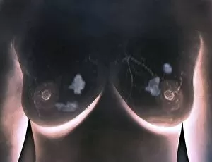
"Unveiling the Hidden Battles: A Glimpse into the World of Oncology" Acute promyelocytic leukaemia, micrograph: Witnessing the intricate battle within, as microscopic warriors fight against acute promyelocytic leukaemia. Brain tumour, 3D-MRI scan: Peering into the depths of complexity, a three-dimensional MRI scan reveals the enigmatic presence of a brain tumour. Breast Cancer. Oil Painting C017 / 1273: Transcending science through artistry, an oil painting captures the essence and resilience in the face of breast cancer. Brain cancer, DTI and 3D CT scans C016 / 6414: Merging technology's prowess with medical expertise, DTI and 3D CT scans expose the intricate web woven by brain cancer. Cardiac lymphoma, MRI scans: Unmasking an elusive foe that lurks within our hearts - cardiac lymphoma unraveled through revealing MRI scans. Throat cancer, X-rays: Illuminating shadows cast upon vocal cords; X-rays shed light on throat cancer's silent invasion. Vaginal cancer cells SEM C014 /0374-0378): Journeying to unseen realms where life hangs in balance - scanning electron microscopy unveils vaginal cancer cells' haunting beauty and danger simultaneously. Dividing liver cancer cell SEM : Witnessing cellular division amidst chaos – scanning electron microscopy exposes dividing liver cancer cells' relentless propagation. Colorectal Cancer Cell SEM : Exploring nature's hidden battlegrounds – scanning electron microscopy uncovers colorectal cancer cells' formidable presence up close. In this captivating journey through oncological landscapes captured by various imaging techniques like micrographs, MRI scans or SEMs we get a glimpse into battles fought at microscopic levels against diseases like acute promyelocytic leukemia or brain tumors.