Fraud Blocker Skip to main content

Optic Nerve Collection

"The Optic Nerve: Unveiling the Intricate Pathways of Vision" Step into the fascinating world of the optic nerve

Background imageOptic Nerve Collection: Rene Descartes' idea of vision

Rene Descartes' idea of vision
5310106 Rene Descartes' idea of vision; (add.info.: Diagram illustrating Rene Descartes' idea of vision, showing the function of the eye, optic nerve, and brain)

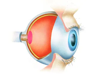
Background imageOptic Nerve Collection: Cross section illustration of anatomy of human eye showing use of optic nerve

Cross section illustration of anatomy of human eye showing use of optic nerve
Anatomy, Biomedical Illustration, Close-Up, Cross Section, Eyelash, Fovea, Human Eye, Iris, No People, Optic Nerve, Part Of, Physiology, Pupil, Sensory Perception, Side View, Single Object

Background imageOptic Nerve Collection: Digital illustration of how human brain uses blindsight to visualize moving tennis ball

Digital illustration of how human brain uses blindsight to visualize moving tennis ball
arrow sign, biomedical illustration, blindsight, brain, connection, digitally generated, direction, human eye, no people, optic nerve, physiology, sensory perception, studio shot, tennis ball

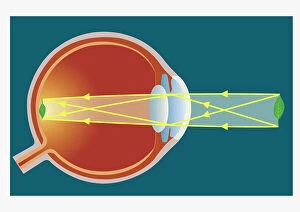
Background imageOptic Nerve Collection: Digital illustration of human eye transforming images made from light into electrical impulses

Digital illustration of human eye transforming images made from light into electrical impulses
arrow sign, biomedical illustration, blue background, connection, cornea, cross section, digitally generated, direction, eyesight, full frame, horizontal, human eye, iris, lens, no people

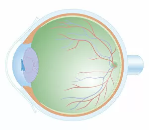
Background imageOptic Nerve Collection: Normal anatomy of the eye in cross section

Normal anatomy of the eye in cross section
Horizontal, Vision, Detail, Anatomy, Diagram, Muscle, Illustration, Lens, Iris, Medical, Eyeball, Biology, Lateral, Cut Out, White Background, Eye, Artwork, Cross Section, Color Image, Close Up

Background imageOptic Nerve Collection: Descartes idea of the hydraulic action of the nerves, 1692

Descartes idea of the hydraulic action of the nerves, 1692. French philosopher Rene Descartes (1596-1650) believed nerves were hollow, provided with valves

Background imageOptic Nerve Collection: Rene Descartes idea of vision, showing the function of the eye, optic nerve and brain, 1692

Rene Descartes idea of vision, showing the function of the eye, optic nerve and brain, 1692. From Opera Philosophica by Rene Descartes. (Frankfurt-am-Main, 1692)

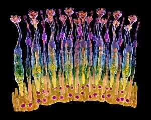
Background imageOptic Nerve Collection: Eye, rods and cones of retina, artwork C017 / 7791

Eye, rods and cones of retina, artwork C017 / 7791
Photograph of an human eye and illustration of rods and cones in the retina. The retina, inside the eye, contains a light-sensitive membranous layer of cells

Background imageOptic Nerve Collection: Retinal implant, artwork

Retinal implant, artwork
Retinal implant. Computer artwork of a biomedical chip (square, centre right) implanted just under the surface of the retina in a human eye

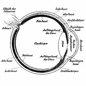
Background imageOptic Nerve Collection: Human eye, published in 1898

Human eye, published in 1898
Antique illustration of a Human eye, published in 1898

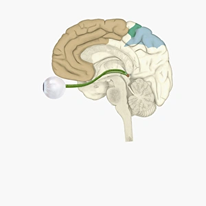
Background imageOptic Nerve Collection: Digital illustration of various areas of cortex in human brain receiving input from sense organs

Digital illustration of various areas of cortex in human brain receiving input from sense organs

Background imageOptic Nerve Collection: Cross section biomedical illustration of anatomy of human eye

Cross section biomedical illustration of anatomy of human eye

Background imageOptic Nerve Collection: Illustration of human eye with muscles, blood vessels and nerves

Illustration of human eye with muscles, blood vessels and nerves
Medicine: Human anatomy, eye. Muscles, blood vessels and nerves

Background imageOptic Nerve Collection: Eye retina F008 / 0713

Eye retina F008 / 0713
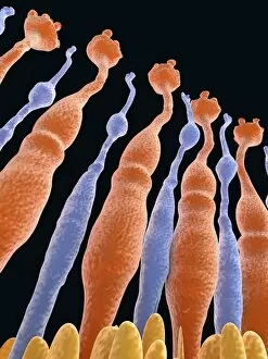
The retina, inside the eye, contains a light-sensitive membranous layer of cells. These are specialized nerve cells: elongated rods (flower like shapes) and cone-tipped cells, that produce vision

Background imageOptic Nerve Collection: Eye retina, rod and cone F008 / 0719

Eye retina, rod and cone F008 / 0719
The retina, inside the eye, contains a light-sensitive membranous layer of cells. These are specialized nerve cells: elongated rods (flower like shape) and cone-tipped cells, that produce vision

Background imageOptic Nerve Collection: Eye retina F008 / 0714

Eye retina F008 / 0714
The retina, inside the eye, contains a light-sensitive membranous layer of cells. These are specialized nerve cells: elongated rods (flower like shapes) and cone-tipped cells, that produce vision

Background imageOptic Nerve Collection: Eye retina F008 / 0712

Eye retina F008 / 0712
The retina, inside the eye, contains a light-sensitive membranous layer of cells. These are specialized nerve cells: elongated rods (flower like shapes) and cone-tipped cells, that produce vision

Background imageOptic Nerve Collection: Eye retina F008 / 0715

Eye retina F008 / 0715
The retina, inside the eye, contains a light-sensitive membranous layer of cells. These are specialized nerve cells: elongated rods (flower like shapes) and cone-tipped cells, that produce vision

Background imageOptic Nerve Collection: Eye retina F008 / 0717

Eye retina F008 / 0717
The retina, inside the eye, contains a light-sensitive membranous layer of cells. These are specialized nerve cells: elongated rods (flower like shapes) and cone-tipped cells, that produce vision

Background imageOptic Nerve Collection: Eye retina F008 / 0711

Eye retina F008 / 0711
The retina, inside the eye, contains a light-sensitive membranous layer of cells. These are specialized nerve cells: elongated rods (flower like shapes) and cone-tipped cells, that produce vision

Background imageOptic Nerve Collection: Eye retina F008 / 0718

Eye retina F008 / 0718
The retina, inside the eye, contains a light-sensitive membranous layer of cells. These are specialized nerve cells: elongated rods (flower like shapes) and cone-tipped cells, that produce vision

Background imageOptic Nerve Collection: Eye retina F008 / 0720

Eye retina F008 / 0720
The retina, inside the eye, contains a light-sensitive membranous layer of cells. These are specialized nerve cells: elongated rods (flower like shapes) and cone-tipped cells, that produce vision

Background imageOptic Nerve Collection: Eye retina, rod and cone F008 / 0716

Eye retina, rod and cone F008 / 0716
The retina, inside the eye, contains a light-sensitive membranous layer of cells. These are specialized nerve cells: elongated rods (flower like shape) and cone-tipped cells, that produce vision

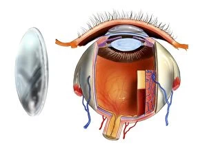
Background imageOptic Nerve Collection: Contact lens and eye anatomy, artwork C016 / 6572

Contact lens and eye anatomy, artwork C016 / 6572
Contact lens and eye anatomy. Artwork of a contact lens (left) and the anatomy of an eye (right) with a contact lens placed on the corneal surface (upper right)

Background imageOptic Nerve Collection: Evolution of the eye, artwork

Evolution of the eye, artwork
Evolution of the eye, computer artwork. It is thought that the eye originated as a patch of light-sensitive cells (left) on the organisms surface

Background imageOptic Nerve Collection: Retina of the eye, light micrograph C016 / 0528

Retina of the eye, light micrograph C016 / 0528
Retina of the eye. Light micrograph of a section through the retina from a human eye. From top down: nerve fibres of the optic nerve and a blood vessel; several layers of neurons (nerve cells)

Background imageOptic Nerve Collection: Bernhard von Gudden, German anatomist C018 / 7084

Bernhard von Gudden, German anatomist C018 / 7084
Bernhard von Gudden (1824-1886). 1885 engraving of the German microanatomist and psychiatrist Johann Bernhard Aloys von Gudden

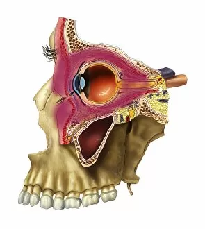
Background imageOptic Nerve Collection: Plegmon eye inflammation, artwork

Plegmon eye inflammation, artwork
Plegmon eye inflammation. Artwork of a sagittal section through a human eye and the surrounding structures, in a case of a subcutaneous (below the skin) phlegmon inflammation of an eye cavity

Background imageOptic Nerve Collection: Eye anatomy, artwork C016 / 8742

Eye anatomy, artwork C016 / 8742
Eye anatomy, artwork. The eye is seen in cutaway format from the side. Structures shown include the lens (light blue, lower left), its attachment points, the iris

Background imageOptic Nerve Collection: Central nervous system, artwork

Central nervous system, artwork
Central nervous system. Computer artwork showing the brain, cranial nerves and the top portion of the spinal cord. At centre is the pituitary gland (small, spherical), which secretes hormones

Background imageOptic Nerve Collection: Eye retina C017 / 7794

Eye retina C017 / 7794
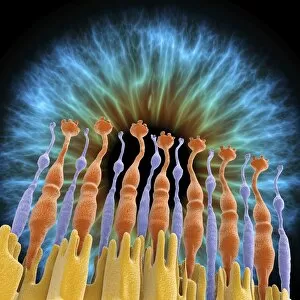
The retina, inside the eye, contains a light-sensitive membranous layer of cells. These are specialized nerve cells: elongated rods (flower like shapes) and cone-tipped cells, that produce vision

Background imageOptic Nerve Collection: Eye retina C017 / 7793

Eye retina C017 / 7793
The retina, inside the eye, contains a light-sensitive membranous layer of cells. These are specialized nerve cells: elongated rods (flower like shapes) and cone-tipped cells, that produce vision

Background imageOptic Nerve Collection: Eye retina C017 / 7792

Eye retina C017 / 7792
The retina, inside the eye, contains a light-sensitive membranous layer of cells. These are specialized nerve cells: elongated rods (flower like shapes) and cone-tipped cells, that produce vision

Background imageOptic Nerve Collection: Eye retina and iris C017 / 7789

Eye retina and iris C017 / 7789
The retina, inside the eye, contains a light-sensitive membranous layer of cells. These are specialized nerve cells: elongated rods (flower like shapes) and cone-tipped cells, that produce vision

Background imageOptic Nerve Collection: Eye retina C017 / 7790

Eye retina C017 / 7790
The retina, inside the eye, contains a light-sensitive membranous layer of cells. These are specialized nerve cells: elongated rods (flower like shapes) and cone-tipped cells, that produce vision

Background imageOptic Nerve Collection: Eye retina and iris C017 / 7788

Eye retina and iris C017 / 7788
The retina, inside the eye, contains a light-sensitive membranous layer of cells. These are specialized nerve cells: elongated rods (flower like shapes) and cone-tipped cells, that produce vision

Background imageOptic Nerve Collection: Eye retina and iris C017 / 7785

Eye retina and iris C017 / 7785
The retina, inside the eye, contains a light-sensitive membranous layer of cells. These are specialized nerve cells: elongated rods (flower like shapes) and cone-tipped cells, that produce vision

Background imageOptic Nerve Collection: Eye retina C017 / 7786

Eye retina C017 / 7786
The retina, inside the eye, contains a light-sensitive membranous layer of cells. These are specialized nerve cells: elongated rods (flower like shapes) and cone-tipped cells, that produce vision

Background imageOptic Nerve Collection: Eye retina C017 / 7784

Eye retina C017 / 7784
The retina, inside the eye, contains a light-sensitive membranous layer of cells. These are specialized nerve cells: elongated rods (flower like shapes) and cone-tipped cells, that produce vision

Background imageOptic Nerve Collection: Eye retina C017 / 7787

Eye retina C017 / 7787
The retina, inside the eye, contains a light-sensitive membranous layer of cells. These are specialized nerve cells: elongated rods (flower like shapes) and cone-tipped cells, that produce vision

Background imageOptic Nerve Collection: Eye retina C017 / 7783

Eye retina C017 / 7783
The retina, inside the eye, contains a light-sensitive membranous layer of cells. These are specialized nerve cells: elongated rods (flower like shapes) and cone-tipped cells, that produce vision

Background imageOptic Nerve Collection: Brain and eye anatomy, artwork C013 / 4664

Brain and eye anatomy, artwork C013 / 4664
Brain and eye anatomy. Computer artwork of the brain from below, with the front of the brain and the eyeballs (white, one at right sectioned) at top

Background imageOptic Nerve Collection: Head and neck anatomy, artwork

Head and neck anatomy, artwork
Head and neck anatomy. Computer artwork of a human head and neck with the overlying tissues removed to show the brain (pink) within a sectioned skull that includes the teeth (white) of the jaws

Background imageOptic Nerve Collection: Optic nerve, light micrograph

Optic nerve, light micrograph
Optic nerve. Coloured light micrograph of a section through the optic nerve. The optic nerve (bright pink) collects impulses from the light sensitive cells of the retina and relays them to the brain

Background imageOptic Nerve Collection: Eye retina

Eye retina
Structure of the retina. Computer artwork showing the structure of the eye retina. The retina is a thin tissue layer on the inner eye responsible for sight. Light strikes from the top

Background imageOptic Nerve Collection: Computer artwork of the structure of the retina

Computer artwork of the structure of the retina
Structure of the retina. Computer artwork showing the structure of the eye retina. The retina is a thin tissue layer on the inner eye responsible for sight. Light strikes from the top

Background imageOptic Nerve Collection: Visible Human Project: sectioned male head

Visible Human Project: sectioned male head

Background imageOptic Nerve Collection: Rapid eye movement, conceptual artwork

Rapid eye movement, conceptual artwork
Rapid eye movement. Conceptual computer artwork representing a saccade, the fast and simultaneous movement of both eyes in the same direction


All Professionally Made to Order for Quick Shipping

"The Optic Nerve: Unveiling the Intricate Pathways of Vision" Step into the fascinating world of the optic nerve, where intricate networks and delicate structures come together to create our sense of sight. Through a combination of scientific illustrations, artwork, and historical references, we embark on a journey that reveals the wonders hidden within this vital pathway. Retina blood vessels captured under an SEM (Scanning Electron Microscope) showcase their intricate patterns, emphasizing their crucial role in nourishing the retina. Head and neck anatomy artwork provides a broader context for understanding how the optic nerve connects with various regions of our body. Delving deeper into its composition, SEM images reveal optic nerve fibers in all their complexity. These microscopic threads transmit visual information from our eyes to the brain at astonishing speeds. A cross-sectional view unveils normal eye anatomy while highlighting key components such as rods and cones found within the retina. Drawing inspiration from history, we explore Rene Descartes' groundbreaking ideas on vision dating back to 1692. His concept of hydraulic action in nerves laid foundations for understanding how signals travel through this remarkable system connecting eye and brain. Artwork depicting retinal implants showcases cutting-edge technology aimed at restoring vision for those with impaired sight. This glimpse into modern advancements reminds us of humanity's relentless pursuit to unlock new possibilities within our own bodies. A captivating illustration published in 1898 takes us back in time when scientists were just beginning to unravel the mysteries behind human ocular function. The intricacies depicted serve as testament to centuries-long fascination with understanding how we perceive light. Finally, digital illustrations highlight different areas of cortex receiving input from sense organs like eyes – reminding us that vision is not solely confined to one isolated structure but rather an integrated process involving multiple regions within our extraordinary brains. Exploring these diverse perspectives allows us to appreciate both past discoveries and present innovations surrounding the optic nerve.