Fraud Blocker Skip to main content

Pathology Collection (page 8)

"Exploring the Intricate World of Pathology: Unveiling the Mysteries Within" Delving into the realm of pathology, we encounter a fascinating array of scientific wonders

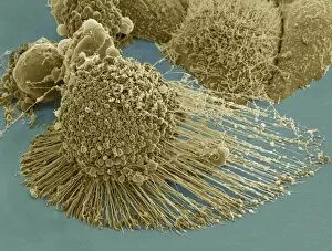
Background imagePathology Collection: Scanning electron micrograph of an apoptotic HeLa cell

Scanning electron micrograph of an apoptotic HeLa cell. Zeiss Merlin HR-SEM

Background imagePathology Collection: Microscopic view of hookworm

Microscopic view of hookworm. The hookworm is a parasitic nematode that lives in the small intestine of its host, which may be a mammal such as a dog, cat, or human

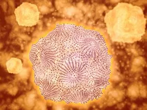
Background imagePathology Collection: Microscopic view of Canine Parvovirus

Microscopic view of Canine Parvovirus. Canine Parvovirus is a highly contagious viral disease that is one of the most common causes of diarrhea in dogs under 6 months of age

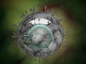
Background imagePathology Collection: Microscopic view of HIV virus

Microscopic view of HIV virus

Background imagePathology Collection: Influenza virus, glassy look

Influenza virus, glassy look

Background imagePathology Collection: Conceptual image of common bacteria

Conceptual image of common bacteria

Background imagePathology Collection: The Manner of Dissecting the Pestilential Body (engraving) (b / w photo)

The Manner of Dissecting the Pestilential Body (engraving) (b / w photo)
XJF135242 The Manner of Dissecting the Pestilential Body (engraving) (b/w photo) by English School, (17th century); Private Collection; English, out of copyright

Background imagePathology Collection: VIRCHOW TITLE-PAGE, 1858. Title-page of Rudolph Virchows Die Cellularpathologie
VIRCHOW TITLE-PAGE, 1858. Title-page of Rudolph Virchows Die Cellularpathologie, 1858

Background imagePathology Collection: USS COMFORT, c1919. The pathology laboratory aboard the hospital ship USS Comfort

USS COMFORT, c1919. The pathology laboratory aboard the hospital ship USS Comfort. Photograph, c1919

Background imagePathology Collection: Rudolph Virchow (1821-1902) German pathologist and founder of cell pathology. In

Rudolph Virchow (1821-1902) German pathologist and founder of cell pathology. In later life he turned to anthropology and archaeology and collaborated with Schliemann on the excavations at Troy

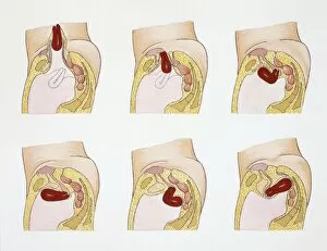
Background imagePathology Collection: Medical illustration of Obstetrics with Internal female reproductive system

Medical illustration of Obstetrics with Internal female reproductive system, Abnormal uterus positions, drawing
Medicine - Anatomy - Obstetrics - Internal female reproductive system - Abnormal uterus positions. Drawing

Background imagePathology Collection: Medical illustration of Digestive system, Intestinal stenosis

Medical illustration of Digestive system, Intestinal stenosis: examples of most common pathologies, drawing
Medicine - Pathology - Digestive system - Intestinal stenosis: examples of most common pathologies. Drawing

Background imagePathology Collection: Malaria, Plasmodium vivax in blood under microscope

Malaria, Plasmodium vivax in blood under microscope
Medicine - Human Histology - Pathology - Malaria - Plasmodium vivax in blood under microscope

Background imagePathology Collection: Pathology, Human Digestive System, Liver, Hepatic cirrhosis, Hepatic tissue destruction under

Pathology, Human Digestive System, Liver, Hepatic cirrhosis, Hepatic tissue destruction under microscope
Medicine - Human Histology - Pathology - Digestive system - Liver - Hepatic cirrhosis - Hepatic tissue destruction under microscope

Background imagePathology Collection: Human Digestive System, Liver, Microscopic view of alcohol-induced damage of hepatic tissue

Human Digestive System, Liver, Microscopic view of alcohol-induced damage of hepatic tissue
Medicine - Human Histology - Pathology - Digestive system - Liver - Microscopic view of alcohol-induced damage of hepatic tissue

Background imagePathology Collection: Atherosclerosis, drawing

Atherosclerosis, drawing
Medicine - Pathology - Atherosclerosis, drawing

Background imagePathology Collection: X-ray of arthritic knee

X-ray of arthritic knee
Medicine - Anatomy - Pathology - Diagnostic Imaging - X-ray of arthritic knee

Background imagePathology Collection: Illustration showing adenoma of parathyroid gland

Illustration showing adenoma of parathyroid gland
Medicine: Human anatomy, pathology, adenoma of the parathyroid gland. Drawing

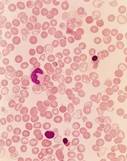
Background imagePathology Collection: Medicine - anatomy - cardio-circulatory system - blood smear with sickle-shaped red blood cells

Medicine - anatomy - cardio-circulatory system - blood smear with sickle-shaped red blood cells
Medicine - Anatomy - Cardio-circulatory system - Blood smear with sickle-shaped red blood cells, typical of Mediterranean anemia or Cooleys disease, at the microscope

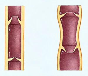
Background imagePathology Collection: Two illustrations showing the flow of blood in a healthy vein and a varicose vein

Two illustrations showing the flow of blood in a healthy vein and a varicose vein

Background imagePathology Collection: Two Pink Shrimp (Malacostracans), close up

Two Pink Shrimp (Malacostracans), close up

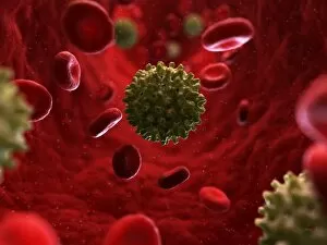
Background imagePathology Collection: Hepatitis B infection, artwork

Hepatitis B infection, artwork
Hepatitis B infection. Computer artwork of Hepatitis B virus particles in the bloodstream

Background imagePathology Collection: Polio infection, artwork

Polio infection, artwork
Polio infection. Computer artwork of human polio virus particles in the bloodstream

Background imagePathology Collection: Foot and mouth infection, artwork

Foot and mouth infection, artwork
Foot and mouth infection. Computer artwork of foot and mouth virus particles in the bloodstream

Background imagePathology Collection: HPV infection, artwork

HPV infection, artwork
HPV infection. Computer artwork of human papillomavirus particles in the bloodstream

Background imagePathology Collection: Hepatitis E infection, artwork

Hepatitis E infection, artwork
Hepatitis E infection. Computer artwork of Hepatitis E virus particles in the bloodstream

Background imagePathology Collection: Norovirus infection, artwork

Norovirus infection, artwork
Norovirus infection. Computer artwork of norovirus particles in the bloodstream

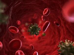
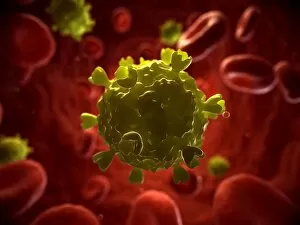
Background imagePathology Collection: HIV infection, artwork

HIV infection, artwork
HIV infection. Computer artwork of HIV particles in the bloodstream

Background imagePathology Collection: Viruses, conceptual artwork

Viruses, conceptual artwork
Viruses, conceptual computer artwork

Background imagePathology Collection: Duret haemorrhages C016 / 8919

Duret haemorrhages C016 / 8919
Duret haemorrhages. Gross specimen of a section through a brain stem showing Duret haemorrhages (bleeding, dark areas). These haemorrhages are most often caused by raised intracranial pressure

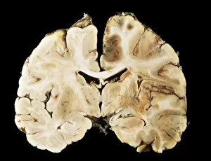
Background imagePathology Collection: Brain injury C016 / 8921

Brain injury C016 / 8921
Brain injury. Gross specimen of a section through a brain showing swelling and haemorrhaging (brown areas) in one hemisphere (right)

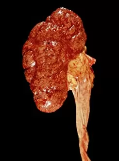
Background imagePathology Collection: Inflamed kidney

Inflamed kidney
Inflamed and swollen kidney, gross specimen. This is a case of chronic nephritis, an inflammation of the kidney. Nephritis is often caused by a bacterial infection

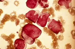
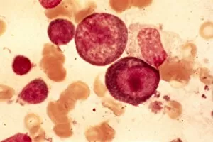
Background imagePathology Collection: Acute myeloid leukaemia, micrograph

Acute myeloid leukaemia, micrograph
Acute myeloid leukaemia. Light micrograph of blood cells from bone marrow in a case of acute myeloid leukaemia. These cells include a monocytoid myeloblast stem cell

Background imagePathology Collection: Elliptocytosis, light micrograph

Elliptocytosis, light micrograph
Elliptocytosis. Light micrograph of red blood cells in a case of elliptocytosis. Red blood cells (erythrocytes) carry oxygen and carbon dioxide to and from body tissues

Background imagePathology Collection: Cystine in bone marrow, light micrograph

Cystine in bone marrow, light micrograph
Cystine in bone marrow. Light micrograph of crystals of cystine among blood cells in a sample of bone marrow. Cystine is an amino acid that can form crystals in urine

Background imagePathology Collection: Pernicious anaemia, light micrograph

Pernicious anaemia, light micrograph
Pernicious anaemia. Light micrograph of megaloblast blood cells from bone marrow in a case of pernicious anaemia, also known as Biermers anaemia

Background imagePathology Collection: Blood cell cancer, light micrograph

Blood cell cancer, light micrograph
Blood cell cancer. Light micrograph of blood cells from a lymphatic ganglion in a case of a blood cancer of a mixed cell type

Background imagePathology Collection: Infectious mononucleosis, micrograph

Infectious mononucleosis, micrograph
Infectious mononucleosis. Light micrograph of blood cells obtained from an inflamed lymphatic ganglion in a case of infectious mononucleosis (glandular fever)

Background imagePathology Collection: Lymphoblastic sarcoma, light micrograph

Lymphoblastic sarcoma, light micrograph
Lymphoblastic sarcoma. Light micrograph of blood cells from bone marrow in a case of lymphoblastic sarcoma. These small lymphoblasts (dark red) characteristically have conspicuous nucleoli

Background imagePathology Collection: Multiple myeloma, light micrograph

Multiple myeloma, light micrograph
Multiple myeloma. Light micrograph of dystrophy (abnormality) of plasmocyte blood cells from bone marrow in a case of multiple myeloma, also known as Kahlers disease or plasma cell myeloma

Background imagePathology Collection: Lymphoplasmacytic lymphoma, micrograph

Lymphoplasmacytic lymphoma, micrograph
Lymphoplasmacytic lymphoma. Light micrograph of a mixture of small lymphocytes and plasma cells from bone marrow in a case of lymphoplasmacytic lymphoma, also known as Waldenstroms macroglobulinemia

Background imagePathology Collection: Hairy cell leukaemia, light micrograph

Hairy cell leukaemia, light micrograph
Hairy cell leukaemia. Light micrograph of blood cells in a case of hairy cell leukaemia. This sample, treated with May-Grunwald-Giemsa (MGG) stain

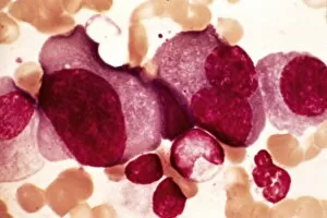
Background imagePathology Collection: Acute erythroid leukaemia, micrograph

Acute erythroid leukaemia, micrograph
Acute erythroid leukaemia. Light micrograph of blood cells from bone marrow in a case of acute erythroid leukaemia. The cells include dystrophic (degenerated) erythroblasts (nuclei stained dark red)

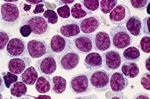
Background imagePathology Collection: Acute lymphoblastic leukaemia, micrograph

Acute lymphoblastic leukaemia, micrograph
Acute lymphoblastic leukaemia. Light micrograph of blood cells from bone marrow in a case of acute lymphoblastic leukaemia. Leukaemia is a cancer where certain blood cells form in excess

Background imagePathology Collection: Malignant histiocytosis, light micrograph

Malignant histiocytosis, light micrograph
Malignant histiocytosis. Light micrograph of blood cells in a case of malignant histiocytosis. Hystiocytosis is an excess number of histiocytes

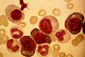
Background imagePathology Collection: Chediak-Higashi syndrome, micrograph

Chediak-Higashi syndrome, micrograph
Chediak-Higashi syndrome. Light micrograph of blood cells in a case of Chediak-Higashi syndrome. The cells include neutrophil white blood cells with abnormal granules


All Professionally Made to Order for Quick Shipping

"Exploring the Intricate World of Pathology: Unveiling the Mysteries Within" Delving into the realm of pathology, we encounter a fascinating array of scientific wonders. From a set of glass eyeballs that have witnessed countless medical marvels to human brain microscope slides revealing intricate neural networks, each specimen holds secrets waiting to be unraveled. Intriguingly, a tobacco hornworm intertwined with its tobacco plant counterpart reminds us of the intricate relationship between organisms and their environment. Similarly, an Alzheimer's brain reveals the devastating effects this disease has on our most precious organ. As we journey through history, we encounter Louis Pasteur, the renowned French microbiologist whose groundbreaking discoveries paved the way for modern medicine. His contributions continue to inspire generations in their pursuit of understanding diseases at their core. Peering through microscopes, we witness captivating views such as that of a microscopic respiratory syncytial virus invading its host or Dohle bodies within blood cells – tiny anomalies holding vital diagnostic clues. The microscopic view of chlamydia serves as a reminder that even seemingly inconspicuous pathogens can wreak havoc on our health. Examining acute promyelocytic leukemia under magnification brings awareness to the battle fought by patients and healthcare professionals alike against this aggressive form of cancer. Meanwhile, glimpsing at sleeping sickness parasites highlights how these minuscule invaders disrupt lives in regions plagued by poverty and limited resources. The sight of liver cirrhosis evokes contemplation about lifestyle choices and their consequences on one's vital organs. In contrast, light micrographs showcasing liver tissue cirrhosis provide insight into cellular changes occurring within this complex disease process. Pathology is not merely confined to textbooks; it is an ever-evolving field where scientists tirelessly strive to decipher nature's enigmatic puzzles. Each slide represents more than just pixels; they hold stories waiting to be told – tales that shed light on diseases afflicting humanity and offer hope for better treatments.