Fraud Blocker Skip to main content

Pathology Collection (page 7)

"Exploring the Intricate World of Pathology: Unveiling the Mysteries Within" Delving into the realm of pathology, we encounter a fascinating array of scientific wonders

Background imagePathology Collection: Microscopic view of human anitbodies with red blood cells

Microscopic view of human anitbodies with red blood cells. Human antibodies are the Y-shaped proteins used by the immune system to defend against foreign objects like bacteria and viruses

Background imagePathology Collection: Virus landing on cell membrane

Virus landing on cell membrane

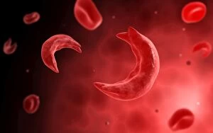
Background imagePathology Collection: Microscopic view of sicke cells causing anemia disease

Microscopic view of sicke cells causing anemia disease

Background imagePathology Collection: Ewings sarcoma locations on the skeleton

Ewings sarcoma locations on the skeleton and detail of tumor on head of femur

Background imagePathology Collection: Microscopic view of rotavirus

Microscopic view of rotavirus. Rotavirus is the most common cause of severe diarrhea among infants and young children. It is a genus of double-stranded RNA virus in the family Reoviridae

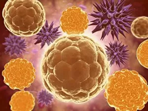
Background imagePathology Collection: Microscopic view of cell and virus

Microscopic view of cell and virus

Background imagePathology Collection: Microscopic view of cancer virus

Microscopic view of cancer virus

Background imagePathology Collection: Microscopic view of a group of Borrelia burgdorferi

Microscopic view of a group of Borrelia burgdorferi, the bacterial agent of Lyme disease transmitted by ticks

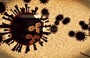
Background imagePathology Collection: A black swarm of H5N1 avian flu viruses are attacked by antibodies

A black swarm of H5N1 avian flu viruses are attacked by antibodies (the three legged elements), which mark the virus for destruction

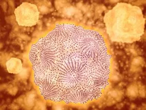
Background imagePathology Collection: Microscopic view of Rubella virus

Microscopic view of Rubella virus
Microscopic view of Rubella. Rubella is an acute, contagious viral infection. While the illness is generally mild in children, it has serious consequences in pregnant women causing fetal death

Background imagePathology Collection: Microscopic view of Legionella pneumophila

Microscopic view of Legionella pneumophila. Legionella pneumophila is a thin, aerobic, pleomorphic, flagellated, non-spore forming, gram-negative bacterium of the genus Legionella

Background imagePathology Collection: Isolated cancer cell Macrophage

Isolated cancer cell Macrophage. Macrophages contribute to tumor growth and progression. Attracted to oxygen-starved (hypoxic) and necrotic tumor cells they promote chronic inflammation

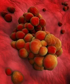
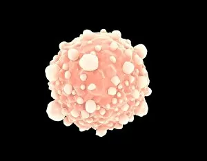
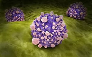
Background imagePathology Collection: Microscopic view of cancer cells

Microscopic view of cancer cells. Cancer occurs when a cells gene mutations make the cell unable to correct DNA damage

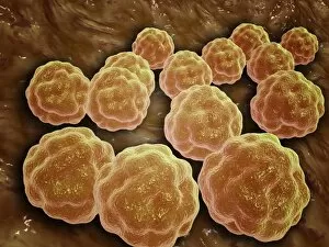
Background imagePathology Collection: Conceptual image of bacteria

Conceptual image of bacteria

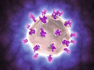
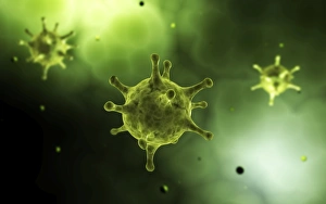
Background imagePathology Collection: Conceptual image of common virus

Conceptual image of common virus

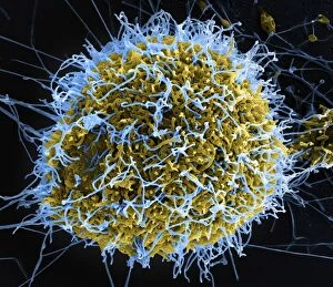
Background imagePathology Collection: Colorized scanning electron micrograph of filamentous Ebola virus particles

Colorized scanning electron micrograph of filamentous Ebola virus particles (blue) budding from a chronically infected VERO E6 cell (yellow-green)

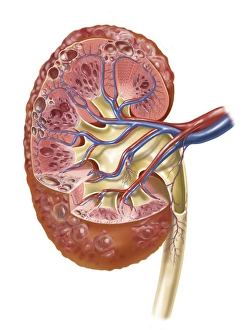
Background imagePathology Collection: Polycystic kidney

Polycystic kidney

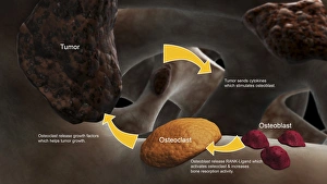
Background imagePathology Collection: Conceptual image illustrating the process of bone metastasis

Conceptual image illustrating the process of bone metastasis

Background imagePathology Collection: Microscopic view of Penicillium expansum

Microscopic view of Penicillium expansum, a mold fungi that effects plants such as apples and similar fruit. This fungus is known as blue mold

Background imagePathology Collection: Conceptual image of the dengue virus

Conceptual image of the dengue virus. The dengue virus is a mosquito-borne RNA virus that causes dengue fever

Background imagePathology Collection: Microscopic view of Yersinia

Microscopic view of Yersinia. Yersinia is a gram-negative, rod-shaped bacteria in the family Enterobacteriaceae

Background imagePathology Collection: Conceptual image of salmonella

Conceptual image of salmonella. Salmonella is a genus of rod-shaped, gram-negative bacteria. There are two main species of Salmonella, Salmonella bongori and Salmonella enterica

Background imagePathology Collection: Microscopic view of a microbe

Microscopic view of a microbe. Microbes are single-cell organisms so tiny that millions can fit into the eye of a needle

Background imagePathology Collection: Microscopic view of tumor

Microscopic view of tumor

Background imagePathology Collection: Microscopic view of bacteria

Microscopic view of bacteria

Background imagePathology Collection: Microscopic view of pancreatic cancer cell

Microscopic view of pancreatic cancer cell

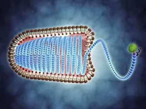
Background imagePathology Collection: Conceptual image of lyssavirus

Conceptual image of lyssavirus. Lyssavirus is a genus of viruses belonging to the family Rhabdoviridae. This group of RNA viruses includes the rabies virus traditionally associated with the disease

Background imagePathology Collection: Conceptual image of a ubiquitous virus

Conceptual image of a ubiquitous virus. A ubiquitous virus is contagious in early childhood through the respiratory tract

Background imagePathology Collection: Microscopic view of Henipavirus

Microscopic view of Henipavirus. Henipavirus is a established group of paramyxoviruses comprising the Hendra virus, Nipah virus and Cedar virus

Background imagePathology Collection: Microscipic view of pancreatic cancer cells

Microscipic view of pancreatic cancer cells

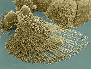
Background imagePathology Collection: Scanning electron micrograph of an apoptotic HeLa cell

Scanning electron micrograph of an apoptotic HeLa cell. Zeiss Merlin HR-SEM

Background imagePathology Collection: Microscopic view of hookworm

Microscopic view of hookworm. The hookworm is a parasitic nematode that lives in the small intestine of its host, which may be a mammal such as a dog, cat, or human

Background imagePathology Collection: Microscopic view of Canine Parvovirus

Microscopic view of Canine Parvovirus. Canine Parvovirus is a highly contagious viral disease that is one of the most common causes of diarrhea in dogs under 6 months of age

Background imagePathology Collection: Microscopic view of HIV virus

Microscopic view of HIV virus

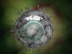
Background imagePathology Collection: Influenza virus, glassy look

Influenza virus, glassy look

Background imagePathology Collection: Conceptual image of common bacteria

Conceptual image of common bacteria

Background imagePathology Collection: The Manner of Dissecting the Pestilential Body (engraving) (b / w photo)

The Manner of Dissecting the Pestilential Body (engraving) (b / w photo)
XJF135242 The Manner of Dissecting the Pestilential Body (engraving) (b/w photo) by English School, (17th century); Private Collection; English, out of copyright

Background imagePathology Collection: VIRCHOW TITLE-PAGE, 1858. Title-page of Rudolph Virchows Die Cellularpathologie

VIRCHOW TITLE-PAGE, 1858. Title-page of Rudolph Virchows Die Cellularpathologie, 1858

Background imagePathology Collection: USS COMFORT, c1919. The pathology laboratory aboard the hospital ship USS Comfort

USS COMFORT, c1919. The pathology laboratory aboard the hospital ship USS Comfort. Photograph, c1919

Background imagePathology Collection: Rudolph Virchow (1821-1902) German pathologist and founder of cell pathology. In

Rudolph Virchow (1821-1902) German pathologist and founder of cell pathology. In later life he turned to anthropology and archaeology and collaborated with Schliemann on the excavations at Troy

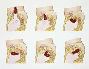
Background imagePathology Collection: Medical illustration of Obstetrics with Internal female reproductive system

Medical illustration of Obstetrics with Internal female reproductive system, Abnormal uterus positions, drawing
Medicine - Anatomy - Obstetrics - Internal female reproductive system - Abnormal uterus positions. Drawing

Background imagePathology Collection: Medical illustration of Digestive system, Intestinal stenosis

Medical illustration of Digestive system, Intestinal stenosis: examples of most common pathologies, drawing
Medicine - Pathology - Digestive system - Intestinal stenosis: examples of most common pathologies. Drawing

Background imagePathology Collection: Malaria, Plasmodium vivax in blood under microscope

Malaria, Plasmodium vivax in blood under microscope
Medicine - Human Histology - Pathology - Malaria - Plasmodium vivax in blood under microscope

Background imagePathology Collection: Pathology, Human Digestive System, Liver, Hepatic cirrhosis, Hepatic tissue destruction under

Pathology, Human Digestive System, Liver, Hepatic cirrhosis, Hepatic tissue destruction under microscope
Medicine - Human Histology - Pathology - Digestive system - Liver - Hepatic cirrhosis - Hepatic tissue destruction under microscope

Background imagePathology Collection: Human Digestive System, Liver, Microscopic view of alcohol-induced damage of hepatic tissue

Human Digestive System, Liver, Microscopic view of alcohol-induced damage of hepatic tissue
Medicine - Human Histology - Pathology - Digestive system - Liver - Microscopic view of alcohol-induced damage of hepatic tissue

Background imagePathology Collection: Atherosclerosis, drawing

Atherosclerosis, drawing
Medicine - Pathology - Atherosclerosis, drawing


All Professionally Made to Order for Quick Shipping

"Exploring the Intricate World of Pathology: Unveiling the Mysteries Within" Delving into the realm of pathology, we encounter a fascinating array of scientific wonders. From a set of glass eyeballs that have witnessed countless medical marvels to human brain microscope slides revealing intricate neural networks, each specimen holds secrets waiting to be unraveled. Intriguingly, a tobacco hornworm intertwined with its tobacco plant counterpart reminds us of the intricate relationship between organisms and their environment. Similarly, an Alzheimer's brain reveals the devastating effects this disease has on our most precious organ. As we journey through history, we encounter Louis Pasteur, the renowned French microbiologist whose groundbreaking discoveries paved the way for modern medicine. His contributions continue to inspire generations in their pursuit of understanding diseases at their core. Peering through microscopes, we witness captivating views such as that of a microscopic respiratory syncytial virus invading its host or Dohle bodies within blood cells – tiny anomalies holding vital diagnostic clues. The microscopic view of chlamydia serves as a reminder that even seemingly inconspicuous pathogens can wreak havoc on our health. Examining acute promyelocytic leukemia under magnification brings awareness to the battle fought by patients and healthcare professionals alike against this aggressive form of cancer. Meanwhile, glimpsing at sleeping sickness parasites highlights how these minuscule invaders disrupt lives in regions plagued by poverty and limited resources. The sight of liver cirrhosis evokes contemplation about lifestyle choices and their consequences on one's vital organs. In contrast, light micrographs showcasing liver tissue cirrhosis provide insight into cellular changes occurring within this complex disease process. Pathology is not merely confined to textbooks; it is an ever-evolving field where scientists tirelessly strive to decipher nature's enigmatic puzzles. Each slide represents more than just pixels; they hold stories waiting to be told – tales that shed light on diseases afflicting humanity and offer hope for better treatments.