
Phagocytosis Collection
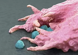
Phagocytosis, the remarkable process of cellular ingestion, plays a crucial role in our immune defense


All Professionally Made to Order for Quick Shipping
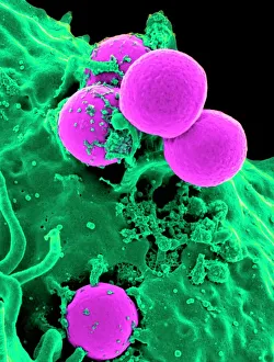
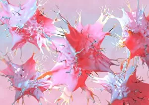
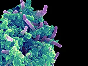
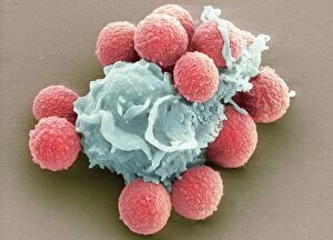
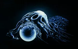
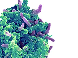
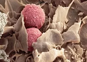
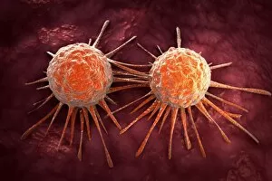
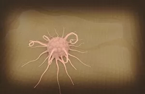
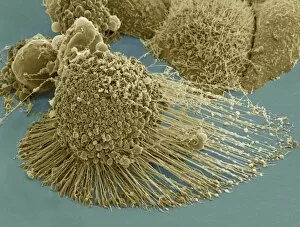
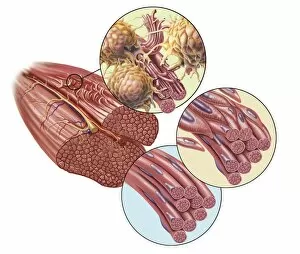
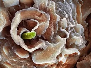
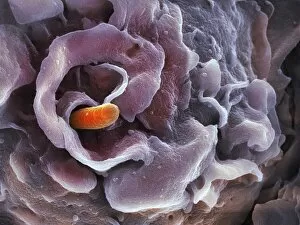
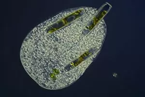
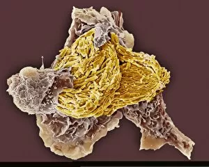
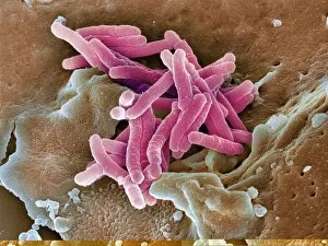
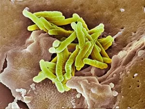
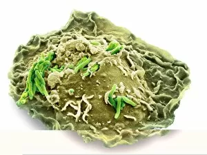
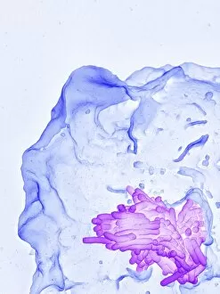
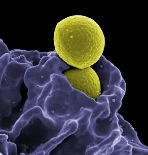
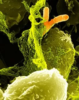
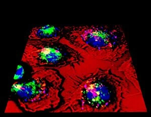
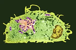
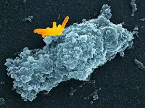
Phagocytosis, the remarkable process of cellular ingestion, plays a crucial role in our immune defense. In this captivating image, we witness the power of neutrophils as they engulf MRSA bacteria, showcasing their ability to devour harmful invaders. The intricate details captured by SEM C018/8596 reveal the intricacies of this life-saving mechanism. Moving on to dendritic cells, an artwork beautifully portrays their pivotal role in phagocytosis. These specialized cells act as sentinels, capturing and presenting antigens to activate other immune cells for a coordinated response against pathogens. Their importance cannot be overstated in maintaining our body's health and vitality. Another striking SEM image showcases a macrophage being invaded by bacteria. This visual representation highlights how these fearless defenders can become targets themselves while fighting off infections. Despite this setback, macrophages remain resilient warriors capable of neutralizing threats through phagocytosis. Intriguingly, phagocytosis extends beyond bacterial invasions; it also encompasses fungal spores' capture and elimination. A mesmerizing SEM image captures the moment when fungal spores are devoured by phagocytes—an extraordinary sight that demonstrates nature's incredible mechanisms at work. The battle against tuberculosis is vividly portrayed through another stunning SEM photograph where a macrophage engulfs TB bacteria with unwavering determination. This powerful display emphasizes how phagocytic cells relentlessly combat infectious agents within our bodies. Not limited to microbial foes alone, macrophages showcase their versatility by engulfing beads—a testament to their adaptability in clearing foreign particles from our system effectively. Multiple sclerosis takes center stage under the scrutiny of an SEM lens—revealing its impact on nerve fibers within our central nervous system. While not directly related to phagocytosis itself, understanding such diseases helps researchers unravel potential therapeutic interventions involving this vital cellular process. Returning once more to dendritic cells, a captivating artwork showcases their elegance and importance in orchestrating immune responses.