Fraud Blocker Skip to main content

Pharynx Collection

The pharynx, also known as the throat, is a vital part of our anatomy

Background imagePharynx Collection: Thyroid anatomy, artwork C013 / 4675

Thyroid anatomy, artwork C013 / 4675
Thyroid anatomy. Computer artwork of the thyroid glands (pink) of the neck, shown in relation to the bodys bones and throat structures (white)

Background imagePharynx Collection: Illustration showing physiology of face and neck of elementary age boy

Illustration showing physiology of face and neck of elementary age boy
6-7 Years, Anatomy, Biomedical Illustration, Caucasian Appearance, Childhood, Close-Up, Development, Elementary Age, Epiglottis, Healthcare and Medicine, Human Face, Human Head Cross Section

Background imagePharynx Collection: Anatomy of male respiratory system and internal organs

Anatomy of male respiratory system and internal organs
Anatomy of male respiratory system, including the mouth, cutaway view. Other internal organs are visible in half tones

Background imagePharynx Collection: Anatomy of female body with internal organs

Anatomy of female body with internal organs

Background imagePharynx Collection: Diseases and disorders of the mouth and throat (colour litho)

Diseases and disorders of the mouth and throat (colour litho)
3097102 Diseases and disorders of the mouth and throat (colour litho) by German School, (19th century); Private Collection; (add.info.: Diseases and disorders of the mouth and throat)

Background imagePharynx Collection: Inflammation chronique du pharynx, Angine granuleuse (colour litho)

Inflammation chronique du pharynx, Angine granuleuse (colour litho)
5210410 Inflammation chronique du pharynx, Angine granuleuse (colour litho) by French School, (19th century); Private Collection; (add.info.: Inflammation chronique du pharynx, Angine granuleuse)

Background imagePharynx Collection: Cross section biomedical illustration of link between the ear, nose and throat

Cross section biomedical illustration of link between the ear, nose and throat

Background imagePharynx Collection: Male anatomy of internal organs

Male anatomy of internal organs, with heart highlighted and rest of organs in half tone

Background imagePharynx Collection: Male skeleton with internal organs on blue background

Male skeleton with internal organs on blue background

Background imagePharynx Collection: Anatomy of human neck

Anatomy of human neck
Neck anatomy showing arteries of pharyngeal region and thyroid, parathyroid glands

Background imagePharynx Collection: Human digestive system, with labels

Human digestive system, with labels

Background imagePharynx Collection: Neck throat anatomy engraving 1866

Neck throat anatomy engraving 1866
Atlas d anatomie descriptive du corps humain C. Bonamy - Paul Broca Victor Masson et Fils Paris 1866

Background imagePharynx Collection: Anatomy of inner ear and sinuses

Anatomy of inner ear and sinuses

Background imagePharynx Collection: Anatomy of female body with internal organs superimposed

Anatomy of female body with internal organs superimposed

Background imagePharynx Collection: Human speech organs, wood engraving, published in 1880

Human speech organs, wood engraving, published in 1880
The Organs of Speech: A) mouth, B) uvula, C) tongue, D) tonsils, E) epiglottis, F) thyroid cartilage, G) arytenoid muscle, H+I) vestibular folds, K) vocal fold, L) larynx, M) hypopharynx, N) trachea

Background imagePharynx Collection: Larynx anatomy, artwork

Larynx anatomy, artwork
Larynx anatomy. Artwork of the anatomy of the human larynx (voicebox) showing the production of vocal sounds (yellow arrows)

Background imagePharynx Collection: Mouth anatomy, artwork

Mouth anatomy, artwork
Mouth anatomy. Artwork of a section through the human head showing the anatomy of the mouth, nose and upper throat. Above the mouth is the nasal cavity (black)

Background imagePharynx Collection: Cross section biomedical illustration of endoscopy of the nose and throat

Cross section biomedical illustration of endoscopy of the nose and throat

Background imagePharynx Collection: Male body standing, with full digestive system superimposed

Male body standing, with full digestive system superimposed
Human male body standing, with full digestive system superimposed

Background imagePharynx Collection: Human digestive system

Human digestive system

Background imagePharynx Collection: Anatomy of a tracheoesophageal fistula

Anatomy of a tracheoesophageal fistula

Background imagePharynx Collection: Anatomy of a childs full digestive system

Anatomy of a childs full digestive system, including mouth and nasal cross section. Other iinternal organs appear in half tone

Background imagePharynx Collection: Male body standing, with full respiratory system superimposed

Male body standing, with full respiratory system superimposed
Human male body standing, with full respiratory system superimposed

Background imagePharynx Collection: The sympathetic nervous system and the organs of fight-or-flight response

The sympathetic nervous system and the organs of fight-or-flight response

Background imagePharynx Collection: Anatomy of a childs full respiratory system

Anatomy of a childs full respiratory system, including mouth and nasal cross section. Other internal organs appear in half tone

Background imagePharynx Collection: Illustration of pharynx, rear view

Illustration of pharynx, rear view
Medicine: Human anatomy, pharynx, back view

Background imagePharynx Collection: Illustration of pharynx, section

Illustration of pharynx, section
Medicine: Human anatomy, pharynx, section. Drawing

Background imagePharynx Collection: Vocal cords, artwork

Vocal cords, artwork
Vocal cords. Computer artwork of a rear view of the larynx (purple/grey), also known as the voice box, and the vocal cords (centre)

Background imagePharynx Collection: Tonsils, artwork

Tonsils, artwork
Tonsils. Artwork of a section through the mouth showing a palatine tonsil. The clear rod points to the tonsillar fossa, a depression that is occupied by a palatine tonsil (round, right)

Background imagePharynx Collection: Oedema of larynx, artwork C016 / 6529

Oedema of larynx, artwork C016 / 6529
Oedema of larynx. Artwork of a transverse (axial) section through a larynx (seen from above) with and without swelling. The bottom image shows a healthy larynx

Background imagePharynx Collection: Oedema of larynx, artwork C016 / 6528

Oedema of larynx, artwork C016 / 6528
Oedema of larynx. Artwork of a vertical (coronal) section through a larynx, showing oedema (swelling caused by fluid accumulation)

Background imagePharynx Collection: Oedema of larynx, artwork C016 / 6530

Oedema of larynx, artwork C016 / 6530
Oedema of larynx. Artwork of sections through a larynx with and without swelling. The bottom image shows a healthy larynx in transverse (axial) section from above

Background imagePharynx Collection: Healthy larynx and thyroid, artwork C014 / 0402

Healthy larynx and thyroid, artwork C014 / 0402
Healthy larynx and thyroid, computer artwork. At centre is the larynx (purple/grey), also known as the voice box, surrounded by the thyroid cartilage (pink).Above the larynx is the hyoid bone (white)

Background imagePharynx Collection: Healthy larynx and thyroid, artwork

Healthy larynx and thyroid, artwork
Healthy larynx and thyroid, computer artwork. At centre is the larynx (purple/grey), also known as the voice box, surrounded by the thyroid cartilage (pink)

Background imagePharynx Collection: Healthy larynx and thyroid, artwork C014 / 0404

Healthy larynx and thyroid, artwork C014 / 0404
Healthy larynx and thyroid, computer artwork. The larynx, also known as the voice box, is purple/grey. It is surrounded by the thyroid cartilage (pink)

Background imagePharynx Collection: Healthy larynx and thyroid, artwork C014 / 0400

Healthy larynx and thyroid, artwork C014 / 0400
Healthy larynx and thyroid, computer artwork. At centre is the larynx (purple/grey), also known as the voice box, surrounded by the thyroid cartilage (pink)

Background imagePharynx Collection: Healthy larynx, artwork C014 / 0403

Healthy larynx, artwork C014 / 0403
Healthy larynx, computer artwork. At centre is the larynx (purple/grey), also known as the voice box, surrounded by the thyroid cartilage (pink)

Background imagePharynx Collection: Throat muscles, 1831 artwork

Throat muscles, 1831 artwork
Throat muscles. Posterior view of the muscles of the pharynx (part of the throat). The pharynx is at centre, with the base of the head at top and the upper part of the chest cavity at bottom

Background imagePharynx Collection: Throat cancer, artwork C013 / 5939

Throat cancer, artwork C013 / 5939
Throat cancer. Cutaway computer artwork of a persons head, showing cancer (yellow) in the pharynx (centre) and oesophagus (lower centre)

Background imagePharynx Collection: Heart and lung anatomy, artwork C016 / 2911

Heart and lung anatomy, artwork C016 / 2911
Heart and lung anatomy. Artwork of an oblique anterior (left-frontal) view of the rib-cage, heart and lungs in a male body. One lung is shown at right

Background imagePharynx Collection: Heart and lung anatomy, artwork C016 / 2909

Heart and lung anatomy, artwork C016 / 2909
Heart and lung anatomy. Artwork of an anterior (frontal) view of the heart and lungs in a male body. One lung is shown at right. The bronchioles (airways) inside the lungs are shown at left

Background imagePharynx Collection: Heart and lung anatomy, artwork C016 / 2904

Heart and lung anatomy, artwork C016 / 2904
Heart and lung anatomy. Artwork of a posterior (from behind) view of the heart and lungs. The heart (mostly obscured) is a hollow muscle that pumps blood around the body and to the lungs

Background imagePharynx Collection: Thyroid anatomy, artwork C013 / 4674

Thyroid anatomy, artwork C013 / 4674
Thyroid anatomy. Computer artwork of the thyroid glands (pink) of the neck, shown in relation to the bodys bones and throat structures (white)

Background imagePharynx Collection: Gem anemone, light micrograph

Gem anemone, light micrograph
Gem anemone. Light micrograph of a transverse section through the body of a gem anemone (Bunodactis verrucosa). This section has passed through the pharynx (stomodaeum) region

Background imagePharynx Collection: Artwork of the nose, mouth and throat in profile

Artwork of the nose, mouth and throat in profile
Nose, mouth and throat. Artwork of a human face in profile showing detail of the mouth, nose and throat. Leading back from the nose (upper left) is the nasal cavity

Background imagePharynx Collection: Nose, mouth and throat

Nose, mouth and throat. Cutaway artwork of the anatomy of the nose, mouth and throat. The large nasal cavity (upper centre) is lined with nerves (yellow) branching from the olfactory bulb (above)

Background imagePharynx Collection: Mouth and throat anatomy, artwork

Mouth and throat anatomy, artwork

Background imagePharynx Collection: Head anatomy, artwork

Head anatomy, artwork
Head anatomy. Artwork showing various aspects of human anatomy in a female head seen from the side. The bones (white) include the lower jaw bone, the bones of the palate, the cheek bones


All Professionally Made to Order for Quick Shipping

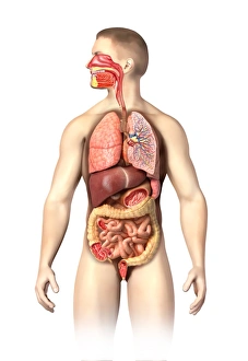
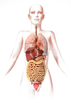
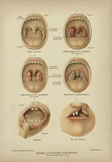
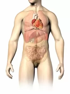
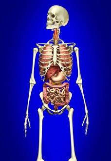
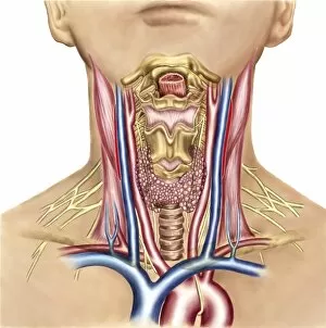
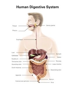
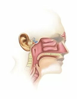
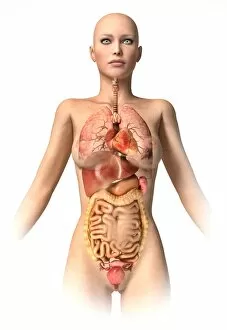
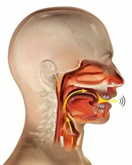
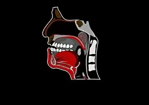
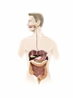
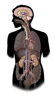
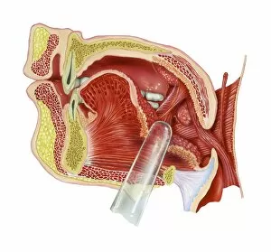
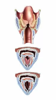
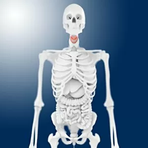
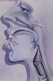
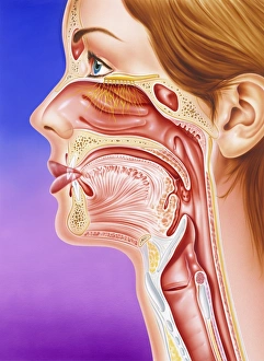
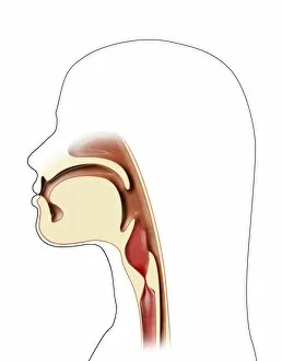
The pharynx, also known as the throat, is a vital part of our anatomy. It plays a crucial role in various bodily functions and is connected to several other organs and systems. When examining the thyroid anatomy, artwork C013 / 4675 provides detailed insights into the structure and function of this important gland located in the neck region. This engraving from 1866 showcases the intricate neck throat anatomy, highlighting how different structures interact within this area. Understanding the respiratory system is essential for comprehending the pharynx's significance. The male respiratory system illustration demonstrates how it connects with internal organs such as lungs and bronchi. Similarly, an anatomical depiction of female body with internal organs sheds light on gender-specific aspects related to this complex network. Exploring diseases and disorders of the mouth and throat becomes easier through color lithographs like "Diseases and Disorders of Mouth and Throat. " These visuals help identify conditions that affect these areas, aiding in diagnosis and treatment. Inflammation chronique du pharynx or angine granuleuse (chronic inflammation or granular sore throat) can be better understood through color lithographs depicting its effects. Such illustrations provide valuable information about symptoms, causes, and potential treatments for these conditions. Sections of the pharynx and larynx offer detailed cross-sectional views that allow us to examine their inner workings more closely. By studying these sections alongside corresponding muscle illustrations, we gain insight into how muscles contribute to swallowing, speaking, breathing processes involving both structures. Biomedical illustrations showcasing links between ear-nose-throat regions highlight their interconnectedness within our bodies' overall functioning. Understanding this connection aids medical professionals when diagnosing issues related to hearing loss or sinus problems affecting both ears' nose-throat region. Anatomy visuals featuring male internal organs provide comprehensive knowledge about organ placement within males specifically while offering comparisons across genders. A skeleton illustration with internal organs against a blue background further enhances our understanding of male anatomy.