
Pistil Collection (page 7)
"Pistil: The Exquisite Beauty of Plant Anatomy Unveiled through Victorian Botanical Illustration and SEM Imagery" Delving into the intricate world of plant anatomy


All Professionally Made to Order for Quick Shipping
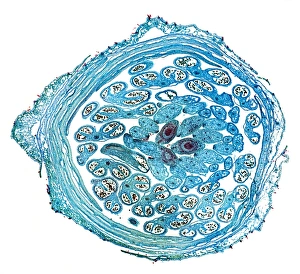
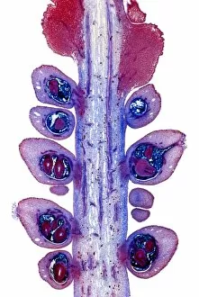
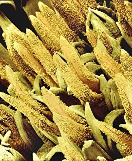
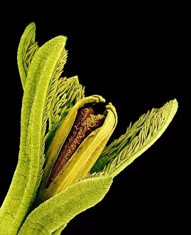
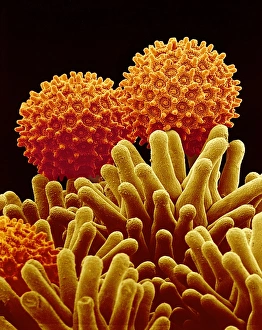
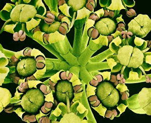
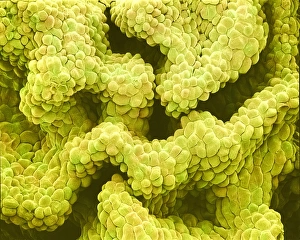
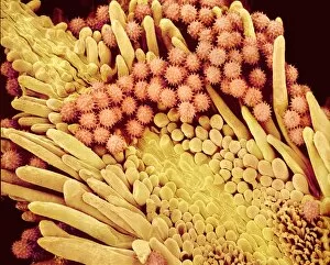
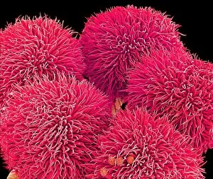
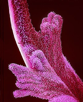
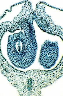
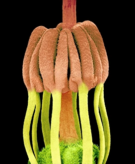
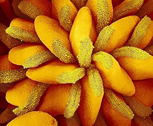
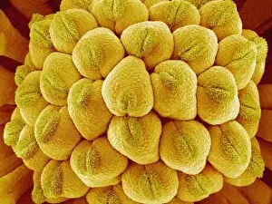
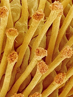
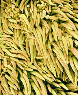
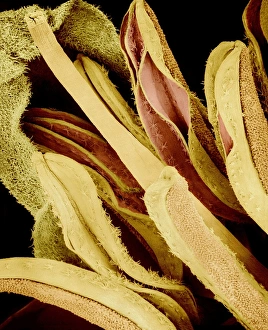
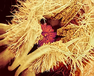
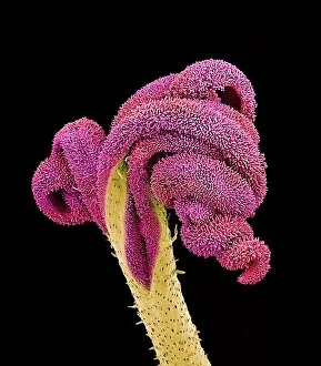
"Pistil: The Exquisite Beauty of Plant Anatomy Unveiled through Victorian Botanical Illustration and SEM Imagery" Delving into the intricate world of plant anatomy, the pistil takes center stage as a captivating subject. Victorian botanical illustrations beautifully capture its delicate structure, showcasing nature's artistry. Intriguingly, the Easter cactus stigma emerges as a focal point in SEM imagery, revealing its mesmerizing texture and detail. Its vibrant colors beckon pollinators to fulfill their crucial role in fertilization. The Pink Azalea Plant portrayed in a Victorian illustration showcases the pistil's elegance amidst lush foliage. A testament to the meticulous observation skills of botanists from yesteryears. SEM images unveil Morning glory pollen up close, displaying its unique shape and surface features. These microscopic grains hold immense significance as they travel through air or insects to reach receptive pistils. A Buttercup flower captured under SEM magnification reveals an enchanting view of its pistil. This image highlights how even seemingly ordinary flowers possess extraordinary beauty when observed at such scale. Pollen tubes of lily pollen showcased by SEM imaging offer insights into this fascinating process called pollination. These slender structures navigate their way towards ovules within the pistil, ensuring successful reproduction. The ethereal dance between pollinators and plants is encapsulated in stunning SEM images capturing moments of pollination itself - a vital step for species survival that relies on precise interaction with each flower's pistil. Amaryllis (Hippeastrum sp. ) stands tall with grace and grandeur while boasting an exquisite display of its reproductive parts under SEM scrutiny. The intricacies within the it can unveiled like never before, inviting admiration for nature's ingenuity. Euphorbia flower reproductive parts take center stage in another compelling SEM image series - offering glimpses into this diverse family's unique adaptations for successful reproduction via their distinctively shaped pistils.