
Planktonic Collection
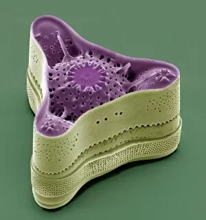
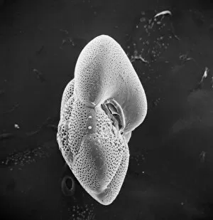
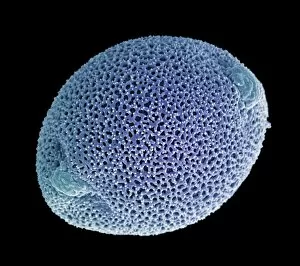
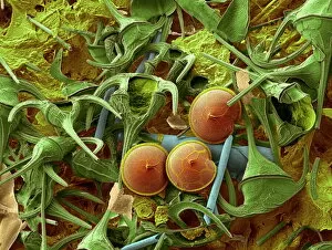
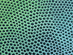
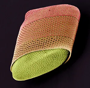
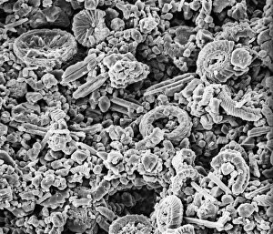
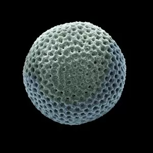
"Exploring the Intricate World Life: Unveiling the Hidden Beauty through SEM Imaging" Diatoms, one of the most fascinating organisms in pond life


All Professionally Made to Order for Quick Shipping
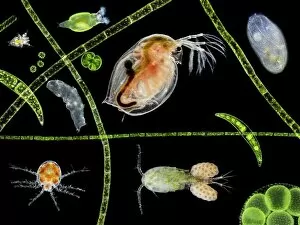
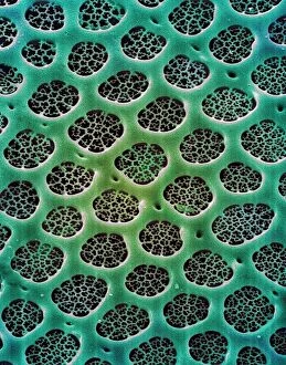
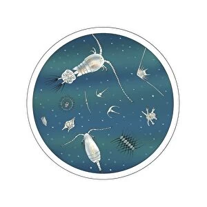
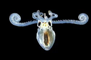
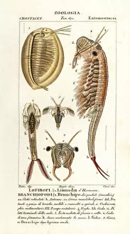
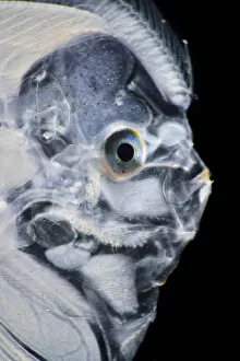
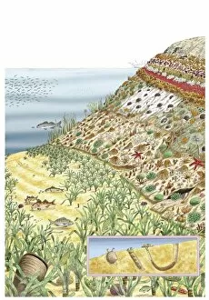
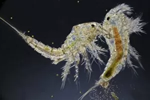
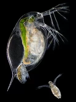
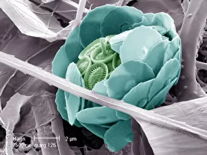
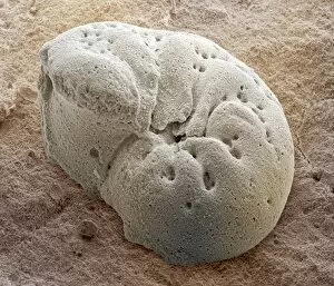
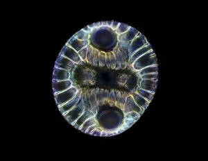
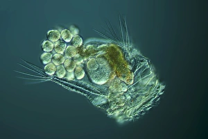
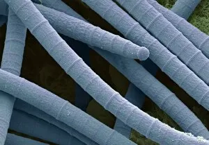
"Exploring the Intricate World Life: Unveiling the Hidden Beauty through SEM Imaging" Diatoms, one of the most fascinating organisms in pond life, reveal their intricate structures under scanning electron microscopy (SEM). The diatom cell wall, composed of silica, showcases a mesmerizing array of patterns and shapes. These microscopic algae play a crucial role in aquatic ecosystems as primary producers. Delving deeper into this captivating realm, we encounter dinoflagellates - another group organisms that exhibit stunning diversity when observed under SEM. Their unique features and complex structures are unveiled through high-resolution imaging techniques. The diatom frustule is an exquisite example of nature's artistry. Its delicate symmetry and intricate designs serve both functional and aesthetic purposes. SEM allows us to appreciate the beauty hidden within these tiny shells. Moving beyond diatoms, we come across foraminifer models - marine protists with elaborate calcium carbonate shells. Examining them using SEM provides insights into their evolutionary adaptations and ecological significance. With each image captured by SEM, we gain a deeper understanding of the complexity and diversity present within planktonic communities. From diatoms to dinoflagellates to foraminifers – these microorganisms shape our planet's ecosystems in ways often unnoticed by human eyes. Through scientific exploration aided by advanced imaging technologies like SEM, we unlock secrets held within these minuscule creatures' world, and is an awe-inspiring journey that reveals not only their structural intricacies but also highlights their vital contributions to Earth's biodiversity and overall environmental health. So next time you gaze upon a tranquil pond or delve into studies on planktonic life, remember the unseen wonders that lie beneath its surface – waiting to be discovered through tools like SEM that bring forth the astonishing beauty found within every diatom shell or algal cell wall.