
Protein Synthesis Collection
Protein synthesis, a fundamental process in all living organisms, plays a crucial role in the growth and maintenance of cells


All Professionally Made to Order for Quick Shipping
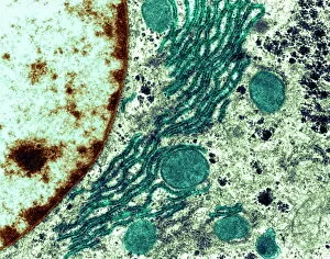
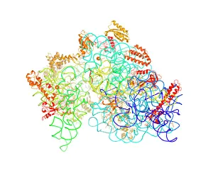
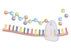
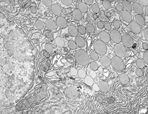
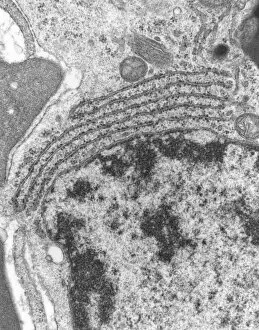
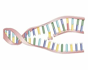
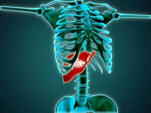
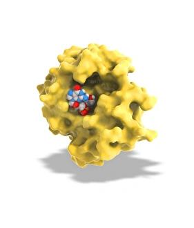
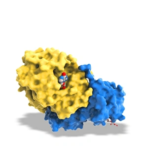
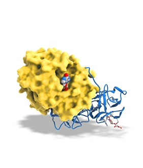
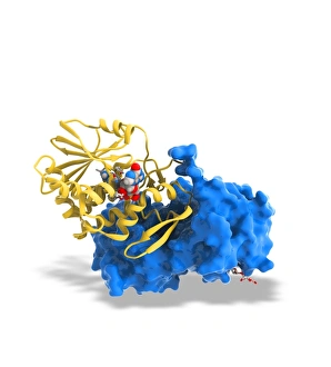
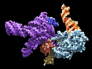
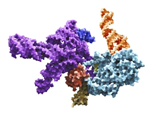
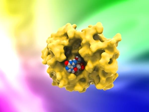
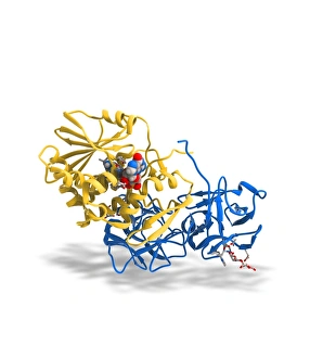
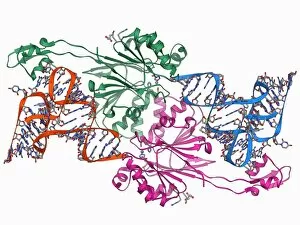
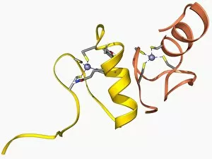
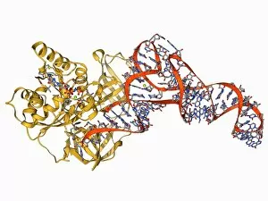
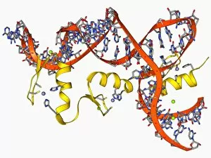
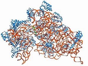
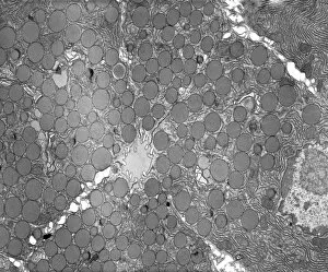
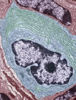
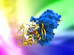
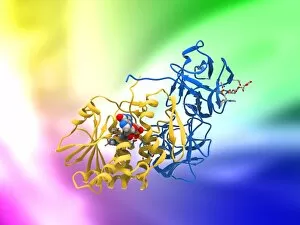
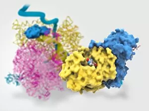
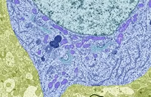
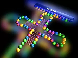
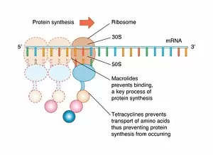
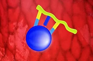
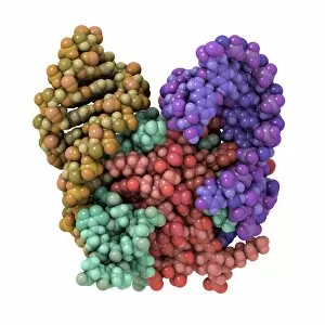
Protein synthesis, a fundamental process in all living organisms, plays a crucial role in the growth and maintenance of cells. This intricate mechanism occurs within the rough endoplasmic reticulum (ER), an organelle responsible for protein production and transportation. Through the lens of a transmission electron microscope (TEM), we can observe the fascinating world of protein synthesis. A bacterial ribosome, resembling a tiny factory, diligently assembles amino acids into complex proteins. In a cross-section biomedical illustration, we witness this remarkable process taking place within DNA – the blueprint of life itself. Moving beyond illustrations, an X-ray view reveals the interconnectedness between protein synthesis and our body's structure. The human skeleton stands tall while relying on proteins produced by liver cells to maintain its strength and integrity. However, not all aspects are beneficial. Ricin A-chain artwork reminds us of its deadly potential when misused or encountered in toxic plants like castor beans. Artworks depicting ricin molecules serve as cautionary reminders about their destructive capabilities if they interfere with essential cellular processes. Returning to explore more positive aspects, let's delve deeper into understanding how proteins are synthesized at a molecular level. Human 80S ribosomes come into focus; these large complexes orchestrate every step involved in assembling amino acids according to genetic instructions encoded by DNA. Intriguingly shaped like clover leaves carrying specific amino acids, transfer RNA (tRNA) molecules act as messengers during protein synthesis. They transport each required building block to the growing chain inside ribosomes with precision and accuracy. As we unravel the intricacies surrounding protein synthesis through various visual aids – from TEM images to biomedical illustrations – we gain insight into one of nature's most vital processes that sustains life itself.