Fraud Blocker Skip to main content

Protozoan Collection (page 3)

"Exploring the Microscopic World: Unveiling the Diversity Life" In this captivating image series

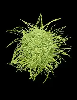
Background imageProtozoan Collection: Heliozoan, SEM C016 / 9024

Heliozoan, SEM C016 / 9024
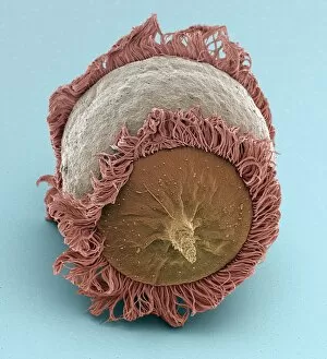
Heliozoan. Coloured scanning electron micrograph (SEM) of a heliozoan freshwater protozoan. This single-celled organism has many projections, known as axopods, radiating from its cell body

Background imageProtozoan Collection: Euplotes protozoa, SEM C016 / 9041

Euplotes protozoa, SEM C016 / 9041
Euplotes protozoa. Coloured scanning electron micrograph (SEM) showing the upperside (bottom) and underside (top) of two Euplotes sp. hypotrich ciliate protozoa

Background imageProtozoan Collection: Climacostomum protozoan, SEM C016 / 9062

Climacostomum protozoan, SEM C016 / 9062
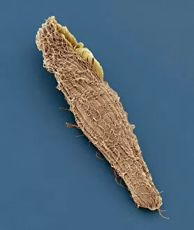
Climacostomum protozoan. Coloured scanning electron micrograph (SEM) of a Climacostomum sp. unicellular ciliate protozoan, showing the cells large oral apparatus (round, upper right)

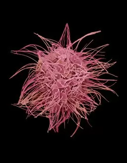
Background imageProtozoan Collection: Heliozoan, SEM C016 / 9023

Heliozoan, SEM C016 / 9023
Heliozoan. Coloured scanning electron micrograph (SEM) of a heliozoan freshwater protozoan. This single-celled organism has many projections, known as axopods, radiating from its cell body

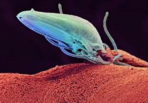
Background imageProtozoan Collection: Giardia protozoan, SEM C016 / 9398

Giardia protozoan, SEM C016 / 9398
Giardia protozoan. Coloured scanning electron micrograph (SEM) of a Giardia sp. trophozoite protozoan (yellow) attached to the lining of a human intestine

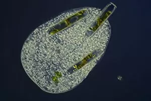
Background imageProtozoan Collection: Ciliate protozoan ingesting algae

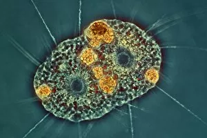
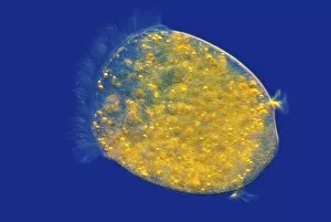
Ciliate protozoan ingesting algae. Darkfield illuminated light micrograph of a ciliate protozoan ingesting green algae (green)

Background imageProtozoan Collection: Tetrahymena protozoan, SEM

Tetrahymena protozoan, SEM
Tetrahymena protozoan. Coloured scanning electron micrograph (SEM) of a Tetrahymena sp. ciliate protozoan. Tetrahymena are free-living freshwater unicellular organisms found in ponds

Background imageProtozoan Collection: Amoeboid protozoa, SEM

Amoeboid protozoa, SEM
Amoeboid protozoa. Differential interference contrast (DIC) micrograph of four Thecamoeba sp. amoebozoa. Amoebozoa are a major group of amoeboid protozoa, motile single-celled organisms

Background imageProtozoan Collection: Ciliate protozoan, light micrograph

Ciliate protozoan, light micrograph
Ciliate protozoan. Darkfield illuminated light micrograph of a ciliate protozoan, showing green algae (green) that it has ingested

Background imageProtozoan Collection: Sleeping sickness parasites, SEM

Sleeping sickness parasites, SEM
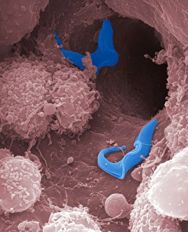
Sleeping sickness parasites. Coloured scanning electron micrograph (SEM) of a Trypanosoma brucei parasites (blue) and white blood cells (pink) in a mouse liver. T
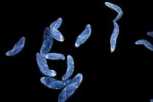
Background imageProtozoan Collection: Sleeping sickness parasite, SEM

Sleeping sickness parasite, SEM
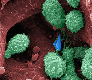
Sleeping sickness parasite. Coloured scanning electron micrograph (SEM) of a Trypanosoma brucei parasite (blue) and a white blood cell (green) in a mouse liver. T

Background imageProtozoan Collection: Amoeba protozoa, SEM

Amoeba protozoa, SEM
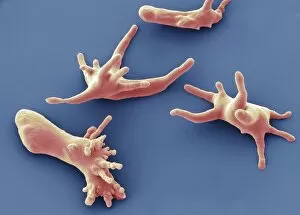
Amoeba protozoa. Coloured scanning electron micrograph (SEM) of four Amoeba sp. protozoa, showing their numerous pseudopodia (cytoplasmic extensions, leg-like)

Background imageProtozoan Collection: Fossil foraminifera necklace C016 / 5995

Fossil foraminifera necklace C016 / 5995
Fossil foraminifera (Alveolina sp.) necklace from Sindh, India. The fossils are from Eocene limestone. Necklace from John Whittakers cabinet. Natural History Museum, London, UK

Background imageProtozoan Collection: Foraminiferan shell, SEM C018 / 0312

Foraminiferan shell, SEM C018 / 0312
Foraminiferan shell, SEM

Background imageProtozoan Collection: Didinium ciliate protozoan, SEM C019 / 0235

Didinium ciliate protozoan, SEM C019 / 0235
Didinium sp. ciliate protozoan, coloured scanning electron micrograph (SEM). This tiny single-celled organism is found in freshwater and marine habitats

Background imageProtozoan Collection: Spirostomum ciliate protozoan, SEM C019 / 0308

Spirostomum ciliate protozoan, SEM C019 / 0308
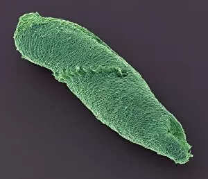
Spirostomum sp. ciliate protozoan, coloured scanning electron micrograph (SEM). These tiny single-celled organisms are found in freshwater and marine habitats

Background imageProtozoan Collection: Blepharisma ciliate protozoan, SEM C019 / 0228

Blepharisma ciliate protozoan, SEM C019 / 0228
Blepharisma ciliate protozoan, coloured scanning electron micrograph (SEM). These tiny single-celled organisms are found in freshwater and marine habitats

Background imageProtozoan Collection: Didinium ciliate protozoan, SEM C019 / 0237

Didinium ciliate protozoan, SEM C019 / 0237
Didinium sp. ciliate protozoan, coloured scanning electron micrograph (SEM). This tiny single-celled organism is found in freshwater and marine habitats

Background imageProtozoan Collection: Spirostomum ciliate protozoan, SEM C019 / 0307

Spirostomum ciliate protozoan, SEM C019 / 0307
Spirostomum sp. ciliate protozoan, coloured scanning electron micrograph (SEM). These tiny single-celled organisms are found in freshwater and marine habitats

Background imageProtozoan Collection: Climacostomum protozoan, SEM C019 / 0243

Climacostomum protozoan, SEM C019 / 0243
Climacostomum sp. ciliate protozoan, coloured scanning electron micrograph (SEM). These tiny single-celled organisms are found in freshwater and marine habitats

Background imageProtozoan Collection: Paramecium sp. protozoa (SEM) C014 / 2148

Paramecium sp. protozoa (SEM) C014 / 2148
Paramecium protozoa. Coloured scanning electron micrograph (SEM) of Paramecium sp. protozoa. These single-celled organisms are aquatic, being found in freshwater habitats

Background imageProtozoan Collection: Radiolarian, SEM C014 / 4862

Radiolarian, SEM C014 / 4862
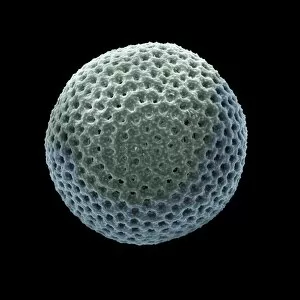
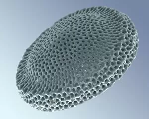
Radiolarian. Coloured scanning electron micrograph (SEM) of the shell or test of a radiolarian. Radiolaria are single-celled protozoans that are found in marine plankton

Background imageProtozoan Collection: Radiolarian, SEM C014 / 4863

Radiolarian, SEM C014 / 4863
Radiolarian. Coloured scanning electron micrograph (SEM) of the shell or test of a radiolarian. Radiolaria are single-celled protozoans that are found in marine plankton

Background imageProtozoan Collection: Radiolaria, light micrograph C014 / 4672

Radiolaria, light micrograph C014 / 4672
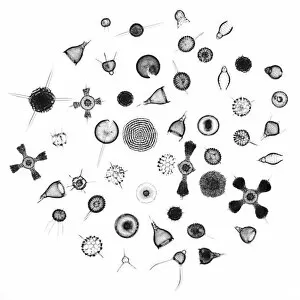
Radiolaria. Collection of light micrographs showing various different species of radiolaria. Radiolaria (or radiolarians) are amoeboid protozoa that produce intricate mineral skeletons

Background imageProtozoan Collection: Ciliate protozoan, light micrograph C014 / 4676

Ciliate protozoan, light micrograph C014 / 4676
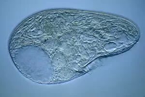
Ciliate protozoan. Differential interference contrast micrograph of a ciliate protozoan, showing the symbiotic green algae (Zoochlorellae, green) contained in vacuoles within

Background imageProtozoan Collection: Ciliate protozoan, light micrograph C014 / 4667

Ciliate protozoan, light micrograph C014 / 4667
Ciliate protozoan. Differential interference contrast micrograph of a ciliate protozoan, showing the symbiotic green algae (Zoochlorellae, green) contained in vacuoles within

Background imageProtozoan Collection: Foraminiferan

Foraminiferan, darkfield light micrograph. Foraminifera are marine single-celled protozoa that construct and inhabit shells composed of several chambers

Background imageProtozoan Collection: Amoeba protozoa, light micrograph C016 / 8607

Amoeba protozoa, light micrograph C016 / 8607
Amoeba protozoa. Interphase contrast light micrograph of two Amoeba sp. protozoa eating each other, showing its numerous pseudopodia (cytoplasmic extensions, leg-like)

Background imageProtozoan Collection: Foraminifera, light micrograph C016 / 8597

Foraminifera, light micrograph C016 / 8597
Foraminifera. Interphase contrast light micrograph of a selection of different foraminifera. Foraminifera are marine single-celled protozoa that construct

Background imageProtozoan Collection: Ciliate protozoan, light micrograph C016 / 8593

Ciliate protozoan, light micrograph C016 / 8593
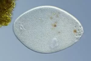
Ciliate protozoan. Differential interference contrast (DIC) light micrograph of a ciliate protozoan. Magnification: x280 when printed 10 centimetres wide

Background imageProtozoan Collection: Hypotrich protozoan, light micrograph C016 / 8592

Hypotrich protozoan, light micrograph C016 / 8592
Hypotrich protozoan. Differential interference contrast (DIC) light micrograph of a Euplotes sp. hypotrich ciliate protozoan. Magnification: x280 when printed 10 centimetres wide

Background imageProtozoan Collection: Hypotrich protozoan, light micrograph C016 / 8590

Hypotrich protozoan, light micrograph C016 / 8590
Hypotrich protozoan. Differential interference contrast (DIC) light micrograph of a Euplotes sp. hypotrich ciliate protozoan. Magnification: x280 when printed 10 centimetres wide

Background imageProtozoan Collection: Ciliate protozoan, light micrograph C016 / 8582

Ciliate protozoan, light micrograph C016 / 8582
Ciliate protozoan. Differential interference contrast (DIC) light micrograph of a ciliate protozoan. Magnification: x280 when printed 10 centimetres wide

Background imageProtozoan Collection: Paramecium bursaria protozoan C016 / 8579

Paramecium bursaria protozoan C016 / 8579
Paramecium bursaria protozoan, phase-contrast light micrograph. This ciliate protozoan inhabits freshwater, where it feeds mainly on bacteria

Background imageProtozoan Collection: Amoeba protozoan, light micrograph C016 / 8578

Amoeba protozoan, light micrograph C016 / 8578
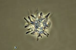
Amoeba protozoan. Phase-contrast light micrograph of an Amoeba sp. protozoan, showing its numerous pseudopodia (cytoplasmic extensions, leg-like)

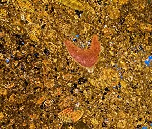
Background imageProtozoan Collection: Fossil-bearing rock, polarised LM C017 / 8483

Fossil-bearing rock, polarised LM C017 / 8483
Fossil-bearing rock, polarised light micrograph. This thin slice from a limestone rock shows various Nummulite fossils. It is a type known as Nummulitie limestone

Background imageProtozoan Collection: Spirostomum protozoa C017 / 8347

Spirostomum protozoa C017 / 8347
Spirostomum protozoa. This large, elongated, single-celled creature has tiny, hair-like structures (cilia) along the length of its body

Background imageProtozoan Collection: Fossilised foraminiferan, SEM C015 / 5772

Fossilised foraminiferan, SEM C015 / 5772
Fossilised foraminiferan. Scanning electron micrograph (SEM) of a section through the fossilised shell of a foraminiferan in a sample of limestone

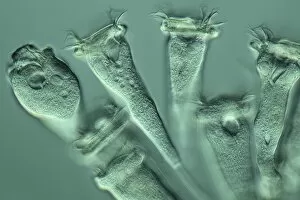
Background imageProtozoan Collection: Vorticella protozoan, light micrograph

Vorticella protozoan, light micrograph
Vorticella protozoan. Rheinberg illuminated light micrograph of a Vorticella sp. ciliate protozoan. This single-celled organism consists of a bell-shaped head

Background imageProtozoan Collection: Paramecium bursaria protozoan, light micr

Paramecium bursaria protozoan, light micr
Paramecium bursaria protozoan, differential interference contrast light micrograph. This ciliate protozoan inhabits freshwater, where it feeds mainly on bacteria

Background imageProtozoan Collection: Blepharisma protozoan, light micrograph

Blepharisma protozoan, light micrograph
Blepharisma protozoan. Differential interference contrast micrograph of a Blepharisma japonicum protozoan. These ciliate protozoa move by beating their cilia

Background imageProtozoan Collection: Blepharisma protozoa, light micrograph

Blepharisma protozoa, light micrograph
Blepharisma protozoa. Differential interference contrast micrograph of Blepharisma japonicum protozoa. These ciliate protozoa move by beating their cilia

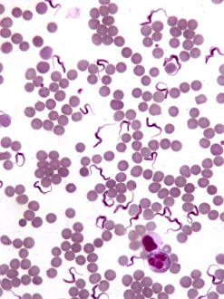
Background imageProtozoan Collection: Trypanosomes in blood smear, SEM C016 / 5783

Trypanosomes in blood smear, SEM C016 / 5783
Parasitic protozoan. Coloured scanning electron micrograph (SEM) of trypanosome protozoa (Trypanosoma sp.) in a blood smear

Background imageProtozoan Collection: Malaria drug dispensing, World War II C016 / 4303

Malaria drug dispensing, World War II C016 / 4303
Malaria drug dispensing. Close-up of the antimalarial drug atabrine being distributed during World War II (1939-1945) according to a checklist

Background imageProtozoan Collection: Malaria drug warning, World War II C016 / 4301

Malaria drug warning, World War II C016 / 4301
Malaria drug warning. Skulls on a warning sign at a US Army hospital during World War II (1939-1945). Also known as quinacrine and mepacrine

Background imageProtozoan Collection: Vorticella protozoa, light micrograph C016 / 3042

Vorticella protozoa, light micrograph C016 / 3042
Vorticella protozoa. Differential interference contrast (DIC) light micrograph of Vorticella sp. ciliate protozoa. These single-celled organisms consist of a bell-shaped head

Background imageProtozoan Collection: Vorticella protozoan, light micrograph C016 / 3043

Vorticella protozoan, light micrograph C016 / 3043
Vorticella protozoan. Differential interference contrast (DIC) light micrograph of a Vorticella sp. ciliate protozoan. This single-celled organism consists of a bell-shaped head

Background imageProtozoan Collection: Vorticella protozoa, light micrograph C016 / 3041

Vorticella protozoa, light micrograph C016 / 3041
Vorticella protozoa. Differential interference contrast (DIC) light micrograph of Vorticella sp. ciliate protozoa. These single-celled organisms consist of a bell-shaped head


All Professionally Made to Order for Quick Shipping

"Exploring the Microscopic World: Unveiling the Diversity Life" In this captivating image series, we delve into the fascinating realm of protozoa – a diverse group of single-celled organisms that inhabit our planet. From ancient calcareous phytoplankton fossils to intricate SEM images, prepare to be amazed by their incredible forms and functions. Starting with a glimpse into the past, we encounter an exquisite fossilized specimen of calcareous phytoplankton. Preserved in time, it offers us a window into Earth's history and evolution. Moving forward, our attention is captured by an artistic representation of Trypanosome protozoan – a notorious parasite responsible for causing sleeping sickness. Its mesmerizing artwork reminds us of both its beauty and danger lurking within nature. Shifting gears towards human health concerns, we come across Plasmodium sp. , the malarial parasite that has plagued humanity for centuries. This microscopic culprit serves as a stark reminder of the ongoing battle against malaria worldwide. Protozoa are known for their unique feeding strategies; some scavenge particles and microorganisms like bacteria while others absorb nutrients from their surroundings. Their adaptability is showcased through stunning SEM images featuring diatoms - intricately patterned unicellular algae - and Acrosphaera radiolarian - delicate marine organisms with intricate skeletal structures. The exploration continues as we encounter another menacing parasite responsible for mouse malaria. Through high-resolution SEM imagery, its complex morphology comes to life before our eyes. Diving deeper into this hidden world reveals Oxytricha ciliate protozoan – an organism characterized by its hair-like projections called cilia. The detailed SEM image showcases its extraordinary structure in vivid detail. Finally, we marvel at the intricate design of Foraminiferan tests (shells) captured using scanning electron microscopy (SEM). These tiny shells serve as protective homes for these remarkable creatures living in aquatic environments.