Fraud Blocker Skip to main content

Radiograph Collection (page 8)

"Unveiling the Hidden World: Exploring Radiographs Beyond the Ordinary" Step into a captivating realm where radiographs reveal secrets hidden beneath our very eyes

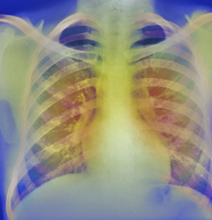
Background imageRadiograph Collection: Heart, chest X-ray

Heart, chest X-ray

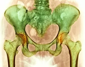
Background imageRadiograph Collection: Healthy hip bones, X-ray

Healthy hip bones, X-ray
Healthy hip bones. Coloured X-ray of the pelvis of a 49 year old woman, showing bones of the lower spine, hips and thighs

Background imageRadiograph Collection: Jaws of a shark

Jaws of a shark, coloured X-ray

Background imageRadiograph Collection: Osteoarthritis of hip joints, X-ray

Osteoarthritis of hip joints, X-ray
Hip joint osteoarthritis. Coloured X-ray of the pelvis of a 72-year-old woman showing areas of osteoarthritis of the hip joints (orange)

Background imageRadiograph Collection: Breast implant calcification, X-ray

Breast implant calcification, X-ray
Breast implant calcification. Coloured mammogram (breast X-ray) of calcification around the borders of a breast implant (pink) in a womans breast (red) as seen from the side

Background imageRadiograph Collection: Lower leg fracture

Lower leg fracture
MODEL RELEASED. Lower leg fracture. Doctor showing a patient an x-ray of his fractured lower leg

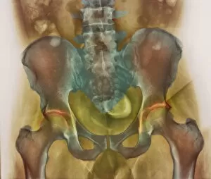
Background imageRadiograph Collection: Osteoarthritic pelvis, X-ray

Osteoarthritic pelvis, X-ray
Osteoarthritis. Coloured X-ray of the pelvis of a 52-year-old man showing areas of osteoarthritis (orange). The two femurs (upper leg bones)

Background imageRadiograph Collection: Lower back pain, X-ray

Lower back pain, X-ray
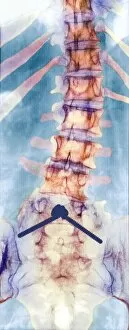
Lower back pain. Conceptual coloured X-ray of the bones of the lower back glowing red in pain. Lower back pain (lumbago) is a very common complaint

Background imageRadiograph Collection: Chronic bronchitis, X-ray

Chronic bronchitis, X-ray
Chronic bronchitis, coloured X-ray. This condition almost exclusively affects smokers. It is often associated with emphysema, which is known as COLD (chronic obstructive lung disease)

Background imageRadiograph Collection: Arthritis of the back, X-ray

Arthritis of the back, X-ray
Arthritis of the back, coloured X-ray. Lumbar arthritis caused by chondrocalcinosis, a condition where crystals of calcium pyrophosphate are deposited in soft tissues

Background imageRadiograph Collection: Pinned curved spine, X-ray

Pinned curved spine, X-ray
Pinned curved spine. Coloured X-ray of bones of the spine and pelvis of a 49 year old woman showing two screws (blue) pinning the lower lumbar vertebra (L5) to the sacrum of the pelvis

Background imageRadiograph Collection: Pinned vertebrae, X-ray

Pinned vertebrae, X-ray
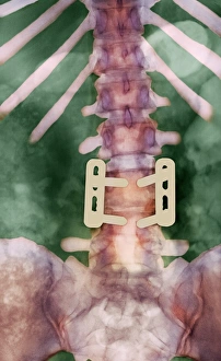
Pinned vertebrae. Coloured X-ray of pins (beige) joining two of the vertebrae (pink) of a 29 year old woman together to immobilise them. Vertebrae are the box-like bones that make up the spine

Background imageRadiograph Collection: Broken upper arm bone, X-ray

Broken upper arm bone, X-ray
Broken upper arm bone. Coloured X-ray of a fracture (yellow) of the upper arm bone (humerus). The neck of the humerus has fractured, separating its head (upper right) from its shaft (below the head)

Background imageRadiograph Collection: Broken arm and dislocated wrist

Broken arm and dislocated wrist. Coloured X-ray showing a fracture (centre) of the radius bone in the forearm and a dislocation of the wrist joint at the end of the ulna bone (centre left)

Background imageRadiograph Collection: Dislocated finger

Dislocated finger

Background imageRadiograph Collection: Finger inflammation, X-ray

Finger inflammation, X-ray
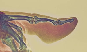
Inflamed finger. Coloured X-ray of a pointing finger, with an inflammatory sore (felon, lower right). The middle finger bone (phalanx, centre) has also been infected and become thinned

Background imageRadiograph Collection: Lower leg fracture, X-ray

Lower leg fracture, X-ray
Lower leg fracture. X-ray of a fracture through the fibula (thinner, left) and tibia or shin (thicker, right) bones of the lower leg

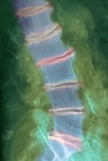
Background imageRadiograph Collection: Spinal osteoporosis, X-ray

Spinal osteoporosis, X-ray
Spinal osteoporosis. Coloured X-ray of the lumbar region of the spine of an elderly female patient with osteoporosis. The bones have lost their density and have become porous and fragile

Background imageRadiograph Collection: Osteoid osteoma, X-ray

Osteoid osteoma, X-ray
Osteoid osteoma. Coloured X-ray of an osteoid osteoma (white, centre left) in the second phalange bone of the middle finger

Background imageRadiograph Collection: Marfans syndrome

Marfans syndrome

Background imageRadiograph Collection: Loss of lung tissue, X-ray

Loss of lung tissue, X-ray
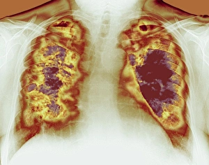
Lung cavities. Coloured chest X-ray showing large cavities (dark red) in the lungs of a patient in a case of Wegeners granulomatosis

Background imageRadiograph Collection: Hiatus hernia, X-ray

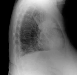
Hiatus hernia, X-ray
Hiatus hernia, coloured X-ray. Side view of a patient suffering from a hiatus hernia, the protrusion of the stomach through the diaphragm into the chest

Background imageRadiograph Collection: Elephantiasis, X-ray

Elephantiasis, X-ray
Elephantiasis. Coloured X-ray showing the lower leg of a patient with elephantiasis, a condition where the lymph vessels (red) become blocked

Background imageRadiograph Collection: Photography

Photography. Conceptual composite image representing photography, with a coloured X-ray of a photographers head and hands using a camera to photograph the Earth

Background imageRadiograph Collection: Arthritic elbow, X-ray

Arthritic elbow, X-ray

Background imageRadiograph Collection: Arthritic hands, X-ray

Arthritic hands, X-ray
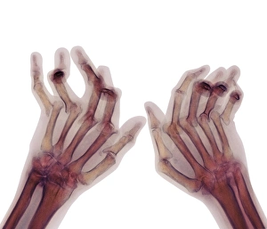
Arthritic hands. Coloured X-ray of the deformed hands of a patient suffering from rheumatoid arthritis. The patients fingers are abnormally bent because of damage to the joints

Background imageRadiograph Collection: Joint disease, CT scan

Joint disease, CT scan

Background imageRadiograph Collection: Arthritic hand bones

Arthritic hand bones
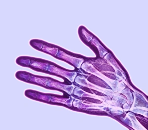
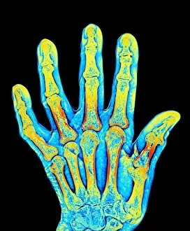
Arthritis. Coloured X-ray of arthritis affecting the joints of the hand. The thumb joint is seen to be both rougher and misaligned compared to a normal joint

Background imageRadiograph Collection: Ankylosing spondylitis X-ray

Ankylosing spondylitis X-ray
Ankylosing spondylitis. Coloured X-ray of the spine of a patient with ankylosing spondylitis, a form of arthritis. This is the lumbar region in the lower back

Background imageRadiograph Collection: Bony spurs in neck, X-ray

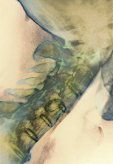
Bony spurs in neck, X-ray
Bony spurs in neck, coloured X-ray. Bony spurs (osteophytes, left) grow from the vertebrae as a normal part of the aging process

Background imageRadiograph Collection: Electric train, X-ray

Electric train, X-ray
Electric train. X-ray showing the mechanism inside an electric toy train

Background imageRadiograph Collection: Toy tin tractor, X-ray

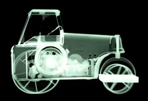
Toy tin tractor, X-ray
Toy tin tractor. X-ray showing the mechanism inside a toy tractor

Background imageRadiograph Collection: Cigarette lighter, simulated X-ray

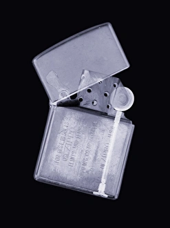
Cigarette lighter, simulated X-ray
Cigarette lighter. Simulated X-ray of a cigarette lighter. The hinged lid of the lighter is slightly open. The main body of the lighter contains a canister of lighter fuel

Background imageRadiograph Collection: Electric light bulbs

Electric light bulbs with a screw fitting, negative image

Background imageRadiograph Collection: Brain scans

Brain scans. Sagittal (sideways) scan through the head, revealing the brain structure. The front of the head is at left. The scan shows the convoluted folds of the outer region

Background imageRadiograph Collection: Pulmonary consolidation, X-ray

Pulmonary consolidation, X-ray
Pulmonary consolidation. X-ray showing that the upper lobe of a patients left lung has become consolidated (filled with pus) due to pneumonia

Background imageRadiograph Collection: Early x-ray of a childs hand

Early x-ray of a childs hand
In 1895 Roentgen discovered a new kind of radiant energy which he named x-rays. On December 22nd 1895 he produced the first human radiograph of his wifes hand

Background imageRadiograph Collection: Early x-ray of a bird

Early x-ray of a bird
In 1895 Roentgen discovered a new kind of radiant energy which he named x-rays. On December 22nd 1895 he produced the first human radiograph of his wifes hand

Background imageRadiograph Collection: 3D hospital scan, conceptual artwork

3D hospital scan, conceptual artwork. Computer artwork of a 3-dimensional image of a human, showing both the muscle and skeletal structure, with X-ray artworks above


All Professionally Made to Order for Quick Shipping

"Unveiling the Hidden World: Exploring Radiographs Beyond the Ordinary" Step into a captivating realm where radiographs reveal secrets hidden beneath our very eyes. From panoramic dental X-rays capturing intricate details of teeth to seahorse skeletons showcasing nature's delicate beauty, these images offer a glimpse into the mesmerizing world of radiography. Delving deeper, we encounter an ordinary foot transformed by an extraordinary X-ray, exposing its inner structure with remarkable precision. A human skull emerges from darkness, unveiling its intricate contours and reminding us of our mortality. Meanwhile, a horse skull stands as a testament to strength and grace in both life and death. But it doesn't stop there - bunions caught on an X-ray remind us that even seemingly small imperfections can leave their mark. In another twist, irregular heartbeats are captured through this powerful imaging technique, shedding light on conditions that affect millions worldwide. Shifting gears towards artistry within science, we witness an MRI-style X-ray revealing a leg adorned in stiletto shoes – merging fashion with medical technology in stunning detail. Parsley springs to life under the gaze of radiographic vision while tulips and gladiolus showcase their vibrant petals through translucent layers. And finally, behold the Oriental stargazer lily – its ethereal beauty enhanced by colored X-rays that transform it into a work of art beyond imagination. Radiographs have transcended mere diagnostic tools; they have become windows into worlds unseen. As we explore these captivating images together, let us marvel at the wonders revealed by this incredible fusion of science and artistry.