Fraud Blocker Skip to main content

Re Production Collection (page 15)

Exploring the wonders of reproduction through time and nature

Background imageRe Production Collection: Pregnant woman, artwork

Pregnant woman, artwork

Background imageRe Production Collection: Mitosis, artwork

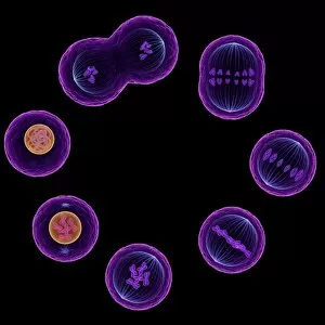
Mitosis, artwork

Background imageRe Production Collection: Human egg cell, artwork

Human egg cell, artwork

Background imageRe Production Collection: Dividing bacteria, artwork

Dividing bacteria, artwork

Background imageRe Production Collection: Uterus lining, SEM

Uterus lining, SEM
Uterus lining, coloured scanning electron micrograph (SEM). At centre is the opening of a secretory gland. The epithelial cells are covered in uniform short microvilli

Background imageRe Production Collection: Cervical lining, SEM

Cervical lining, SEM
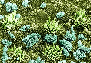
Cervical lining. Coloured scanning electron micrograph (SEM) of microvilli on the lining of the cervix. Magnification: x12000 when printed at 10 centimetres tall

Background imageRe Production Collection: Strawberry pollen, SEM

Strawberry pollen, SEM
Strawberry (Fragaria vesca) pollen grain, coloured scanning electron micrograph (SEM). Magnification: x2000 when printed at 10 centimetres wide

Background imageRe Production Collection: Skimmia pollen, SEM

Skimmia pollen, SEM
Skimmia sp. pollen grain, coloured scanning electron micrograph (SEM). Magnification: x2000 when printed at 10 centimetres wide

Background imageRe Production Collection: Primula pollen, SEM

Primula pollen, SEM
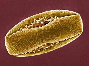
Primrose (Primula sp.) pollen grain, coloured scanning electron micrograph (SEM). Magnification: x2000 when printed at 10 centimetres wide

Background imageRe Production Collection: Epididymis, SEM

Epididymis, SEM
Epididymis. Coloured scanning electron micrograph (SEM) of a freeze fracture through the epididymis. Immature sperm (bright pink at top) are seen in the tubule lumen

Background imageRe Production Collection: Flowering currant pollen, SEM

Flowering currant pollen, SEM
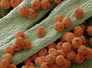
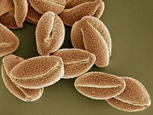
Flowering currant (Ribes sanguineum) pollen grains, coloured scanning electron micrograph (SEM). Magnification: x2000 when printed at 10 centimetres wide

Background imageRe Production Collection: Immature dog sperm, SEM

Immature dog sperm, SEM
Dog sperm. Coloured scanning electron micrograph (SEM) of immature dog sperm. These immature sperm are characterised by a small amount of unshed residual cytoplasm about a third of the way down

Background imageRe Production Collection: Mendelian inheritance

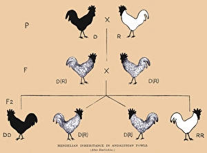
Mendelian inheritance in fowls, as a result of parental genes. P = parents, F & F2 = 1st and 2nd generation, D = dominant, R = recessive

Background imageRe Production Collection: Taq polymerase replicating DNA

Taq polymerase replicating DNA, molecular model. The Taq polymerase is blue, the two strands of DNA are green, and the nucleotide bases are grey (carbon), pink (nitrogen)

Background imageRe Production Collection: Fallopian tube, TEM

Fallopian tube, TEM
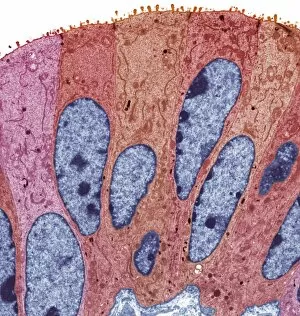
Fallopian tube, coloured transmission electron micrograph (TEM). Section through non-ciliated columnar epithelium from a fallopian tube. The ciliated cells are not shown here

Background imageRe Production Collection: DNA helices over a boys face

DNA helices over a boys face
MODEL RELEASED. DNA molecules superimposed on a boys face. This could represent genetic individuality, or the fact that everyone is a product of the combination of the genes of their father and mother

Background imageRe Production Collection: Chinese hibiscus pollen, SEM

Chinese hibiscus pollen, SEM
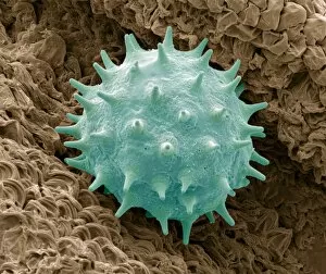
Chinese hibiscus pollen. Coloured scanning electron micrograph (SEM) of a pollen grain from a Chinese hibiscus (Hibiscus rosa-sinensis) flower

Background imageRe Production Collection: Coloured SEM of uterus wall (early proliferation)

Coloured SEM of uterus wall (early proliferation)
Uterus (early proliferation). Coloured scanning electron micrograph (SEM) of the endometrium (lining) of the human uterus during the early proliferative stage of the menstrual cycle

Background imageRe Production Collection: Bellflower pollen, SEM

Bellflower pollen, SEM
Bellflower pollen. Coloured scanning electron micrograph (SEM) of pollen grains from a bellflower (Campanula sp.). Pollen grains are the male gametes (sex cells) of a plant

Background imageRe Production Collection: Fallopian tube cells, SEM

Fallopian tube cells, SEM
Fallopian tube cells. Coloured scanning electron micrograph (SEM) of the surface of a fallopian tube, showing non-ciliated (pink) and ciliated (yellow) cells

Background imageRe Production Collection: Seminal vesicle, light micrograph

Seminal vesicle, light micrograph
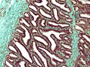
Seminal vesicle. Light micrograph of a section through the seminal vesicle showing the complex mucosal folding.Pseudostratified cuboidal epithelium overlies the loose connective tissue of the lamina

Background imageRe Production Collection: Seminiferous tubules, light micrograph

Seminiferous tubules, light micrograph
Seminiferous tubules. Coloured light micrograph of a section through the testis, showing seminiferous tubules (pink) and leydig cells (yellow). This is the site of sperm production (spermatogenesis)

Background imageRe Production Collection: Epididymis, light micrograph

Epididymis, light micrograph. The lumen of the duct (white) is lined with pseudostratified epithelium (pink), which is made up of columnar cells with elongated nuclei

Background imageRe Production Collection: Blastocyst, artwork

Blastocyst, artwork

Background imageRe Production Collection: Fossilized pollen

Fossilized pollen grains, computer artwork. Pollen grains are reproductive structures produced by flowers. It is rare, but not impossible, for soft structures to be fossilized

Background imageRe Production Collection: Climbing lily pollen, SEM

Climbing lily pollen, SEM
Climbing lily pollen grains. Coloured scanning electron micrograph (SEM) of pollen grains from the climbing lily flower (Gloriosa superba)

Background imageRe Production Collection: Pregnancy timings

Pregnancy timings
MODEL RELEASED. Pregnancy timings. Pregnant woman holding a clock in one hand while supporting her swollen abdomen in the other hand. She is 30 weeks pregnant

Background imageRe Production Collection: Orchid flower (family Orchidaceae)

Orchid flower (family Orchidaceae). Close-up of the lip (lower centre) used to attract pollinators by guiding them to the reproductive parts of the flower

Background imageRe Production Collection: Clematis montana pollen, SEM

Clematis montana pollen, SEM
Clematis montana pollen grains, coloured scanning electron micrograph (SEM). C. montana is a climbing plant that is popular in gardens for its showy flowers

Background imageRe Production Collection: Pregnant woman and son

Pregnant woman and son
MODEL RELEASED. Pregnant woman and son. 4-year-old boy holding and listening to the swollen abdomen of his mother. She is 30 weeks pregnant. A full- term pregnancy lasts between 37 and 42 weeks

Background imageRe Production Collection: Horse chestnut pollen, SEM

Horse chestnut pollen, SEM
Horse chestnut pollen grain. Coloured scanning electron micrograph (SEM) of a pollen grain from a flowering horse chestnut tree (Aesculus hippocastanum)

Background imageRe Production Collection: Pregnant women

Pregnant women
MODEL RELEASED. Pregnant women comparing each others swollen abdomens. They are both about 7 months pregnant. A full-term pregnancy lasts about 9 months (between 37 and 42 weeks)

Background imageRe Production Collection: Pregnancy back pain

Pregnancy back pain
MODEL RELEASED. Pregnant woman holding her painful back as the weight of the unborn child puts strain on her back muscles. She is 30 weeks pregnant. A full-term pregnancy lasts between 37 and 42 weeks

Background imageRe Production Collection: piracy, conceptual artwork

piracy, conceptual artwork
Copyright piracy, conceptual computer artwork

Background imageRe Production Collection: Pregnant woman with birthing ball

Pregnant woman with birthing ball
MODEL RELEASED. Pregnant woman leaning on a large inflatable rubber ball, known as a birthing ball, that is identical to and has a similar function to exercise balls

Background imageRe Production Collection: Pregnancy pains

Pregnancy pains
MODEL RELEASED. Pregnant woman holding her swollen abdomen while wincing in pain. This may be due to compression of internal organs, or movements by the unborn child. The woman is 30 weeks pregnant

Background imageRe Production Collection: Sperm cell, SEM

Sperm cell, SEM
Sperm cell. Coloured scanning electron micrograph (SEM) of a sperm cell in a fallopian tube. Magnification: x400 when printed at 10 centimetres wide

Background imageRe Production Collection: Cave spider, egg sac and spiderlings

Cave spider, egg sac and spiderlings. Cave huntsman spider (Heteropoda) with egg sac and spiderings, on the ceiling of a cave. Large individuals can have legs over 7.5 centimetres long

Background imageRe Production Collection: Cabbage white butterfly caterpillar, SEM

Cabbage white butterfly caterpillar, SEM
Cabbage white butterfly (Pieris brassicae) larva (caterpillar) on the underside of a leaf, coloured scanning electron micrograph (SEM)

Background imageRe Production Collection: Copepod with eggs

Copepod with eggs. Free-swimming copepod carrying a cluster of eggs (red, left) under its tail. Copepods are microscopic aquatic crustaceans found in waters around the world

Background imageRe Production Collection: Copepod

Copepod. Copepods are microscopic aquatic crustaceans found in waters around the world. They may either be free living or parasitic on other organisms

Background imageRe Production Collection: Cabbage white caterpillar hatching, SEM

Cabbage white caterpillar hatching, SEM
Cabbage white butterfly (Pieris brassicae) larva (caterpillar) hatching, coloured scanning electron micrograph (SEM). The eggs are laid in batches of up to 150 on the underside of brassica plant

Background imageRe Production Collection: Flagtail pipefish with eggs

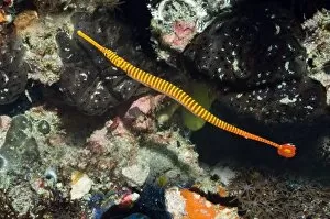
Flagtail pipefish with eggs. Male flagtail pipefish (Dunckerocampus pessuliferus) carrying eggs attached to its underside. Photographed off Rinca Island, Komodo National Park, Indonesia


All Professionally Made to Order for Quick Shipping

Exploring the wonders of reproduction through time and nature. 🌍🔬 From the intricate Catalan Atlas of the 14th century to Da Vinci's innovative crossbow, humans have always sought ways to understand and enhance reproduction. Witness the ancient mating rituals of Tyrannosaurus rex dinosaurs captured in fossils, or marvel at the microscopic beauty of a uterus lining during menstruation under an SEM microscope. Delve into the miracle of life with illustrations showcasing the human placenta or observe English oak acorns as they prepare for their own reproductive journey. Discover nature's secrets with stunning images of geranium anthers and dahlia flower pollen magnified by SEM technology. Follow maple seed flight paths as they embark on their quest for new beginnings, just like Robert Louis Stevenson once wrote about in his captivating works on reproduction. Dive deep into marine life with sea cucumbers, fascinating creatures that possess unique methods of reproduction. And finally, explore the intricate world of plant fertility with mesmerizing SEM images capturing pollen grains in all their glory. Join us on this incredible journey through time and nature as we unravel the mysteries behind 'reproduction'.