
Renal Collection (page 3)
Renal, the intricate masterpiece of our cardiovascular system, has long been celebrated in historical artwork


All Professionally Made to Order for Quick Shipping
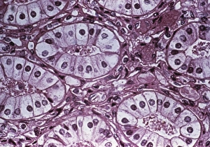
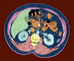
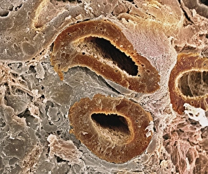
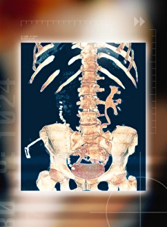
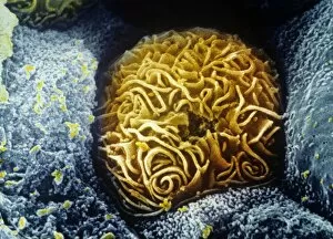
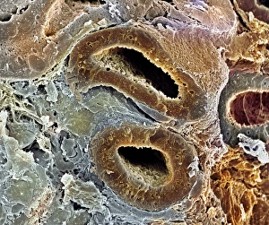
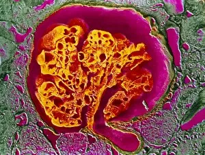
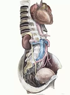
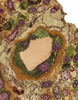
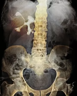
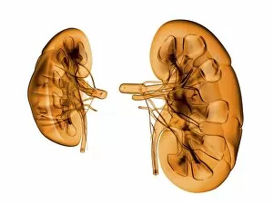
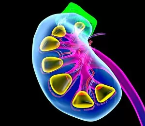
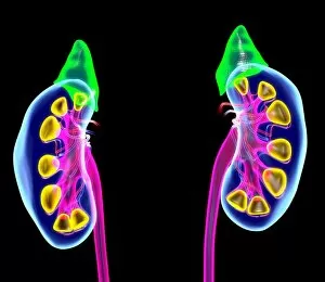
Renal, the intricate masterpiece of our cardiovascular system, has long been celebrated in historical artwork. From ancient paintings to modern sculptures, artists have attempted to capture the essence of this vital organ. When we delve deeper into its structure, we discover kidney tubules in section - a maze-like network responsible for filtering waste and maintaining fluid balance within our bodies. And if one is fortunate enough to possess spare kidneys, they serve as backup guardians of our health. The kidney glomerulus, captured magnificently through scanning electron microscopy (SEM), reveals its intricate web-like structure that allows it to efficiently filter blood and remove toxins. These mesmerizing images remind us of the beauty hidden within our own bodies. Abdominal arteries come alive in an X-ray image (P206 / 0309) showcasing their role in supplying oxygen-rich blood to the renal region. This snapshot highlights the interconnectedness between various components of our body's architecture. Kidney dialysis plays a crucial role for those whose renal function is compromised. It acts as a lifeline by artificially replicating the filtration process performed by healthy kidneys. This medical marvel ensures that individuals can continue living fulfilling lives despite their renal challenges. As we explore more SEM images capturing kidney glomeruli and blood vessels, we are reminded once again of nature's exquisite design. The delicate balance maintained within these structures reflects the harmony required for optimal functioning. Intriguingly juxtaposed with abdominal organs lies this artistic portrayal of kidneys - symbolic representations reminding us how essential they are for overall well-being. They silently work day and night behind-the-scenes performing countless tasks necessary for maintaining homeostasis within our bodies. Renal - not just another word but an embodiment of life-sustaining mechanisms intricately woven into every fiber of human existence. Let us appreciate this remarkable organ that tirelessly serves as a guardian protecting us from harm while quietly orchestrating numerous physiological processes essential for survival.