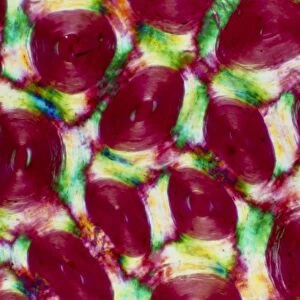
Kidney tubules in section

![]()
Wall Art and Photo Gifts from Science Photo Library
Kidney tubules in section
Kidney tubules. Fluorescent light micrograph of a section through kidney tissue showing numerous tubules (red/green). The tubules are seen in cross-section. Their walls are dark red, with cell nuclei stained blue. The inner lining of the tubules is green. Tubules are part of the filtration system in a kidney. Blood passes through capillaries in the kidney, and fluid and molecules of waste products pass into the tubules. During the passage through the tubules, much of the water is reabsorbed, leaving a concentrated solution of waste products. This drains to the ureters, which pass it to the bladder as urine
Science Photo Library features Science and Medical images including photos and illustrations
Media ID 6450657
© THOMAS DEERINCK, NCMIR/SCIENCE PHOTO LIBRARY
Corpuscle Corpuscles Cross Section Distal Dyed Filtering Filtration Fluorescent Light Micrograph Histological Histology Kidney Kidneys Nephrology Nuclei Nucleus Proximal Renal Stained Tagged Tubule Tubules Urinary System Urine Light Microscope Section Sectioned
EDITORS COMMENTS
This print showcases the intricate structure of kidney tubules, providing a fascinating glimpse into the inner workings of our urinary system. Fluorescent light micrograph technology has beautifully captured numerous tubules in cross-section, revealing their distinct features and functions. The walls of these tubules are depicted in a striking dark red hue, while the cell nuclei appear vividly blue. What truly stands out is the vibrant green inner lining of the tubules, highlighting their crucial role in filtration. As part of the kidney's filtration system, these tubules play a vital role in maintaining our body's balance by removing waste products from our blood. As blood passes through capillaries within the kidneys, fluid and molecules containing waste products enter these remarkable tubular structures. During this journey through the tubules, much of the water is reabsorbed back into circulation while leaving behind a concentrated solution of waste materials. Ultimately, this concentrated solution drains into ureters that transport it to our bladder as urine – an essential process for maintaining overall health and well-being. This visually stunning image not only demonstrates normal anatomy but also serves as a testament to how intricately designed our bodies are at microscopic levels. Captured with precision and artistry by Science Photo Library's fluorescent light micrograph technique, this print offers an educational insight into renal histology and nephrology without mentioning any commercial use or affiliation with any specific company.
MADE IN AUSTRALIA
Safe Shipping with 30 Day Money Back Guarantee
FREE PERSONALISATION*
We are proud to offer a range of customisation features including Personalised Captions, Color Filters and Picture Zoom Tools
SECURE PAYMENTS
We happily accept a wide range of payment options so you can pay for the things you need in the way that is most convenient for you
* Options may vary by product and licensing agreement. Zoomed Pictures can be adjusted in the Cart.