
Schwann Cell Collection
Schwann cells, the unsung heroes of our nervous system, play a vital role in regenerating and myelinating nerve cells


All Professionally Made to Order for Quick Shipping
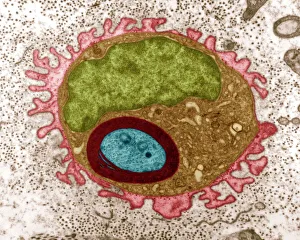
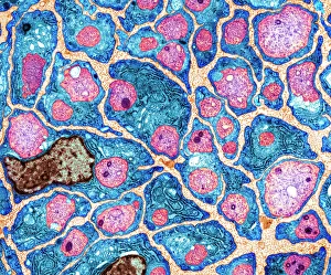
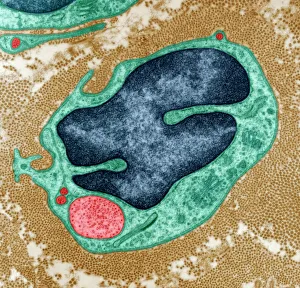
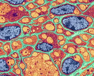
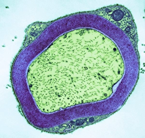
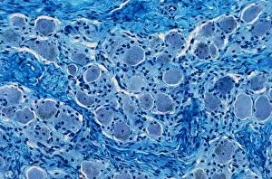
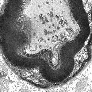
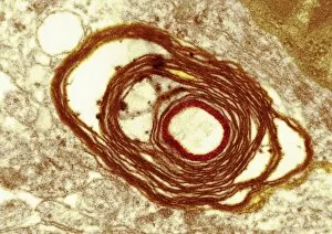
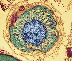
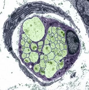
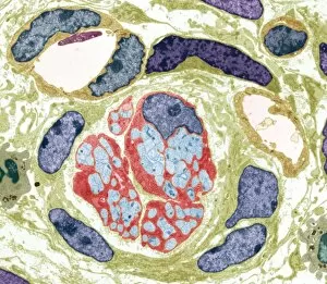
Schwann cells, the unsung heroes of our nervous system, play a vital role in regenerating and myelinating nerve cells. Through the use of transmission electron microscopy (TEM), scientists have been able to unravel the intricate processes involved. In one TEM image, we witness the awe-inspiring sight of a regenerating nerve cell. Schwann cells surround and support these damaged nerves, aiding in their recovery and growth. This remarkable ability highlights their crucial role in repairing our neural pathways. Another TEM image showcases the mesmerizing process of myelination. Schwann cells wrap themselves around nerve fibers, creating a protective layer called myelin. This insulation enhances signal transmission speed and efficiency within our nervous system. Stepping away from TEM imagery, light micrographs offer us glimpses into different aspects biology. In one such image, we observe peripheral nerves with their characteristic branching patterns under a microscope's lens. These networks are responsible for connecting distant parts of our body to transmit sensory information. Moving deeper into the microscopic world, another light micrograph reveals an enchanting view of a nerve ganglion - clusters of neuronal cell bodies surrounded by supportive schwann cells. These ganglia act as relay stations for signals traveling through our nervous system. A closer look at yet another light micrograph unveils beautifully myelinated nerves coursing through tissue like delicate threads interwoven seamlessly together. The presence of this white matter signifies efficient communication between various regions within our body. Returning to TEM images once more brings us face-to-face with intricately detailed views of individual components that make up these complex neural circuits: nerve fibers and nodes. Nerve fiber nodes serve as critical junctions where electrical impulses jump from one node to another along myelinated axons – enabling rapid signal propagation throughout our body. Not all nerves are myelinated though; some remain unmyelinated but still essential for transmitting information accurately across shorter distances.