Fraud Blocker Skip to main content

Stigma Collection (page 5)

"Breaking the Chains: Unraveling the Stigma Surrounding Nature's Wonders" In Saint Joseph of Cupertino's Ecstasy

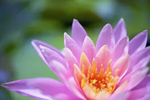
Background imageStigma Collection: Close up of a pink flower in bloom; Hawaii, united states of america

Close up of a pink flower in bloom; Hawaii, united states of america

Background imageStigma Collection: Close Up Of A Hibiscus Flower With Water Droplets; Hawaii, United States Of America

Close Up Of A Hibiscus Flower With Water Droplets; Hawaii, United States Of America

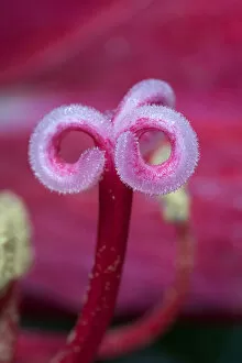
Background imageStigma Collection: Close Up Of Stamen And Filament Of Lily; Quebec, Canada

Close Up Of Stamen And Filament Of Lily; Quebec, Canada

Background imageStigma Collection: Close Up Of A Lily; Quebec, Canada

Close Up Of A Lily; Quebec, Canada

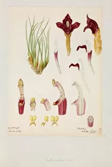
Background imageStigma Collection: Maxillaria subulata

Maxillaria subulata - By Franz Bauer, 1818. Paper, watercolour. Botany Library, Bauer Unit. Unfinished Drawings Monocotyledons 2, Orchidaceae 2, f. 149. Date: 1818

Background imageStigma Collection: The Vision of Saint Francis, ca. 1620, by Carlo Saraceni (15

The Vision of Saint Francis, ca. 1620, by Carlo Saraceni (15
Carlo Saraceni (1580-1620). Italian painter. The Vision of Saint Francis, ca. 1620. Alte Pinakothek. Munich. Germany

Background imageStigma Collection: Francis of Assisi Stigma

Francis of Assisi Stigma
SAINT FRANCIS OF ASSISI displays his stigmata Date: 1182 - 1226

Background imageStigma Collection: USA, California, Death Valley National Park. Detail of a Mojave mound cactus flower

USA, California, Death Valley National Park. Detail of a Mojave mound cactus flower. Credit as: Dennis Flaherty / Jaynes Gallery / DanitaDelimont

Background imageStigma Collection: Life cycle of a flowering plant

Life cycle of a flowering plant

Background imageStigma Collection: Illustration of fertilization of flower, pollen grain containing male nuclei on stigma

Illustration of fertilization of flower, pollen grain containing male nuclei on stigma, male nucleus travelling down pollen tube, female nuclei inside ovule

Background imageStigma Collection: Ms Fr. Fv VI #1 fol. 149r Illustration from the Book of Simple Medicines

Ms Fr. Fv VI #1 fol. 149r Illustration from the Book of Simple Medicines
XJL212064 Ms Fr. Fv VI #1 fol.149r Illustration from the Book of Simple Medicines by Mattheaus Platearius (d.c.1161) c.1470 (vellum) by Testard, Robinet (fl.1470-1523); 26x35.5 cm; National Library

Background imageStigma Collection: USA, California. A flower stigma. Credit as: Christopher Talbot Frank / Jaynes Gallery

USA, California. A flower stigma. Credit as: Christopher Talbot Frank / Jaynes Gallery / DanitaDelimont

Background imageStigma Collection: Picture No. 11072922

Picture No. 11072922
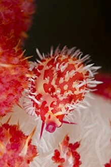
Stigma Egg Cowrie camouflaged on soft coral Nudi Retreat dive site, Lembeh Straits, Sulawesi, Indonesia. Stigma Egg Cowrie

Background imageStigma Collection: Picture No. 10896358

Picture No. 10896358
Parasitic Wasp - Female outside the gall (Glyphomerus stigma) Date:

Background imageStigma Collection: Side view section of a daffodil flower, Narcissus Jetfire, with yellow sepals and orange corona

Side view section of a daffodil flower, Narcissus Jetfire, with yellow sepals and orange corona, ovary, pistil, stigma, filaments and anthers

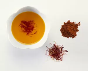
Background imageStigma Collection: Ground saffron

Ground saffron

Background imageStigma Collection: Using paintbrush to move anthers onto stigma, close-up

Using paintbrush to move anthers onto stigma, close-up

Background imageStigma Collection: Hybridising roses, hands brushing anthers of pollinator across stigmas of seed parent, close-up

Hybridising roses, hands brushing anthers of pollinator across stigmas of seed parent, close-up

Background imageStigma Collection: Rubbing anthers of pollen parent over stigma of seed-bearing parent (cross-pollinating a tomato)

Rubbing anthers of pollen parent over stigma of seed-bearing parent (cross-pollinating a tomato), close-up

Background imageStigma Collection: Botany, Dicotyledons, Upper part of style and stigma, illustration

Botany, Dicotyledons, Upper part of style and stigma, illustration
Botany - Dicotyledons - Upper part of style and stigma. Illustration

Background imageStigma Collection: Carnation flower pistil, SEM C016 / 9762

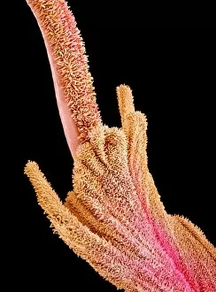
Carnation flower pistil, SEM C016 / 9762
Carnation (Dianthus sp.) pistil, coloured scanning electron micrograph (SEM). Pistils hold a plants female reproductive structures

Background imageStigma Collection: Carnation flower pistil, SEM C016 / 9759

Carnation flower pistil, SEM C016 / 9759
Carnation (Dianthus sp.) pistil, coloured scanning electron micrograph (SEM). Pistils hold a plants female reproductive structures

Background imageStigma Collection: Carnation flower pistil, SEM C016 / 9760

Carnation flower pistil, SEM C016 / 9760
Carnation (Dianthus sp.) pistil, coloured scanning electron micrograph (SEM). Pistils hold a plants female reproductive structures

Background imageStigma Collection: Flowers reproductive structures, artwork

Flowers reproductive structures, artwork
Flowers reproductive structures, computer artwork. At centre is the female part of the flower, the carpel (green), which consists of a stigma (top), style (down centre) and ovary (bottom)

Background imageStigma Collection: Hibiscus flower pistil, SEM C016 / 9758

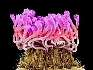
Hibiscus flower pistil, SEM C016 / 9758
Hibiscus flower pistil. Coloured scanning electron micrograph (SEM) of a Hibiscus sp. flower pistil. Pistils hold a plants female reproductive structures

Background imageStigma Collection: Carnation flower pistil, SEM C016 / 9763

Carnation flower pistil, SEM C016 / 9763
Carnation (Dianthus sp.) pistil, coloured scanning electron micrograph (SEM). Pistils hold a plants female reproductive structures

Background imageStigma Collection: Hibiscus flower pistil, SEM C016 / 9757

Hibiscus flower pistil, SEM C016 / 9757
Hibiscus flower pistil. Coloured scanning electron micrograph (SEM) of a Hibiscus sp. flower pistil. Pistils hold a plants female reproductive structures

Background imageStigma Collection: Carnation flower pistil, SEM C016 / 9761

Carnation flower pistil, SEM C016 / 9761
Carnation (Dianthus sp.) pistil, coloured scanning electron micrograph (SEM). Pistils hold a plants female reproductive structures

Background imageStigma Collection: Carnation flower pistil, SEM C016 / 0127

Carnation flower pistil, SEM C016 / 0127
Carnation (Dianthus sp.) pistil, coloured scanning electron micrograph (SEM). Pistils hold a plants female reproductive structures

Background imageStigma Collection: Carnation flower pistil, SEM C016 / 0131

Carnation flower pistil, SEM C016 / 0131
Carnation (Dianthus sp.) pistil, coloured scanning electron micrograph (SEM). Pistils hold a plants female reproductive structures

Background imageStigma Collection: Carnation flower pistil, SEM C016 / 0119

Carnation flower pistil, SEM C016 / 0119
Carnation (Dianthus sp.) pistil, coloured scanning electron micrograph (SEM). Pistils hold a plants female reproductive structures

Background imageStigma Collection: Flower pistils, SEM C016 / 0053

Flower pistils, SEM C016 / 0053
Flower pistils, coloured scanning electron micrograph (SEM). Pistils hold a plants female reproductive structures. They contain one or more carpels, which, in turn, contain a stigma

Background imageStigma Collection: Rose pistil, SEM C015 / 9173

Rose pistil, SEM C015 / 9173
Rose pistil. Coloured scanning electron micrograph (SEM) of a rose (Rosa sp.) flower pistil. Pistils hold a plants female reproductive structures

Background imageStigma Collection: Rose pistil, SEM C015 / 9181

Rose pistil, SEM C015 / 9181
Rose pistil. Coloured scanning electron micrograph (SEM) of a rose (Rosa sp.) flower pistil. Pistils hold a plants female reproductive structures

Background imageStigma Collection: Pea flower pollination, SEM C016 / 0067

Pea flower pollination, SEM C016 / 0067
Pea flower pollination. Coloured scanning electron micrograph (SEM) of pollen grains (ovals) on the stigma of a garden pea flower (Pisum sativum)

Background imageStigma Collection: Flower pistils, SEM C016 / 0055

Flower pistils, SEM C016 / 0055
Flower pistils, coloured scanning electron micrograph (SEM). Pistils hold a plants female reproductive structures. They contain one or more carpels, which, in turn, contain a stigma

Background imageStigma Collection: Flower pistils, SEM C016 / 0054

Flower pistils, SEM C016 / 0054
Flower pistils, coloured scanning electron micrograph (SEM). Pistils hold a plants female reproductive structures. They contain one or more carpels, which, in turn, contain a stigma

Background imageStigma Collection: Flower pistils, SEM C016 / 0057

Flower pistils, SEM C016 / 0057
Flower pistils, coloured scanning electron micrograph (SEM). Pistils hold a plants female reproductive structures. They contain one or more carpels, which, in turn, contain a stigma

Background imageStigma Collection: Carnation flower pistil, SEM C016 / 0129

Carnation flower pistil, SEM C016 / 0129
Carnation (Dianthus sp.) pistil, coloured scanning electron micrograph (SEM). Pistils hold a plants female reproductive structures

Background imageStigma Collection: Flower pistils, SEM C016 / 0058

Flower pistils, SEM C016 / 0058
Flower pistils, coloured scanning electron micrograph (SEM). Pistils hold a plants female reproductive structures. They contain one or more carpels, which, in turn, contain a stigma

Background imageStigma Collection: Pea flower pollination, SEM C016 / 0065

Pea flower pollination, SEM C016 / 0065
Pea flower pollination. Coloured scanning electron micrograph (SEM) of pollen grains (ovals) on the stigma of a garden pea flower (Pisum sativum)

Background imageStigma Collection: Flower pistil, SEM C016 / 0060

Flower pistil, SEM C016 / 0060
Flower pistil, coloured scanning electron micrograph (SEM). Pistils hold a plants female reproductive structures. They contain one or more carpels, which, in turn, contain a stigma

Background imageStigma Collection: Hibiscus flower pistil, SEM C015 / 9160

Hibiscus flower pistil, SEM C015 / 9160
Hibiscus flower pistil. Coloured scanning electron micrograph (SEM) of a Hibiscus sp. flower pistil. Pistils hold a plants female reproductive structures

Background imageStigma Collection: Flower pistils, SEM C016 / 0056

Flower pistils, SEM C016 / 0056
Flower pistils, coloured scanning electron micrograph (SEM). Pistils hold a plants female reproductive structures. They contain one or more carpels, which, in turn, contain a stigma

Background imageStigma Collection: Judas tree flower, SEM C016 / 0061

Judas tree flower, SEM C016 / 0061
Judas tree flower. Coloured scanning electron micrograph (SEM) of the reproductive parts of the flower of the Judas tree, or redbud (Cercis sp.)

Background imageStigma Collection: Flower pistil, SEM C016 / 0059

Flower pistil, SEM C016 / 0059
Flower pistil, coloured scanning electron micrograph (SEM). Pistils hold a plants female reproductive structures. They contain one or more carpels, which, in turn, contain a stigma

Background imageStigma Collection: Pea flower pollination, SEM C016 / 0066

Pea flower pollination, SEM C016 / 0066
Pea flower pollination. Coloured scanning electron micrograph (SEM) of pollen grains (ovals) on the stigma of a garden pea flower (Pisum sativum)

Background imageStigma Collection: Judas tree flower, SEM C016 / 0062

Judas tree flower, SEM C016 / 0062
Judas tree flower. Coloured scanning electron micrograph (SEM) of the reproductive parts of the flower of the Judas tree, or redbud (Cercis sp.)


All Professionally Made to Order for Quick Shipping

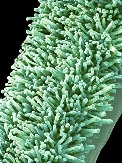
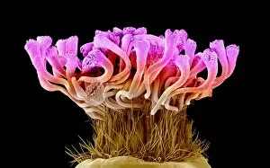
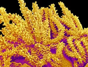
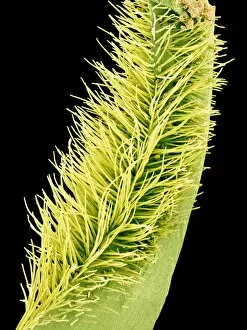
"Breaking the Chains: Unraveling the Stigma Surrounding Nature's Wonders" In Saint Joseph of Cupertino's Ecstasy, we witness a divine connection that transcends human understanding. Just like this masterpiece, stigma in nature holds profound significance. The Methodus plantarum sexalis in sistemate naturae descripta reveals the intricate classification of plants, shedding light on how stigma plays a vital role in their reproductive processes, and is through these delicate structures that life perpetuates itself. Zooming into an Easter cactus stigma under SEM, we discover its mesmerizing beauty up close. The Studio shot captures the essence of a pink tulip flower with such precision that we can almost feel its velvety touch. Morning glory pollen captured by SEM showcases the interplay between stigma and pollen grains—a dance essential for successful pollination. Similarly, Gorse stigma adorned with pollen grains exemplifies nature's harmonious collaboration to ensure survival. A False-colour SEM image of chickweed flower reminds us that even seemingly insignificant flora possess unique stigmas worthy of admiration. Each microscopic detail tells a story waiting to be unraveled. Pollination takes center stage as different species intertwine their destinies through this sacred union. Amaryllis stands tall as Hippeastrum sp. , showcasing vibrant stigmas inviting pollinators to partake in their splendorous feast. Traveling to Greece, we encounter Saffron Crocus—its precious saffron harvested from meticulously hand-picked stigmas—an ancient tradition steeped in culture and history. Lastly, Thale cress flower micrograph serves as a testament to scientific exploration; it invites us into its miniature world where every aspect contributes to our understanding of life's intricacies. Through these captivating glimpses into nature's wonders, let us challenge societal norms and break free from the chains of prejudice surrounding "stigma. " May we recognize its inherent beauty and importance, both in art and the natural world.