
Stomata Collection
"Exploring the Fascinating World of Stomata: Leaf Pores Revealed Through SEM" In the microscopic realm


All Professionally Made to Order for Quick Shipping
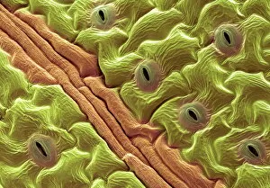
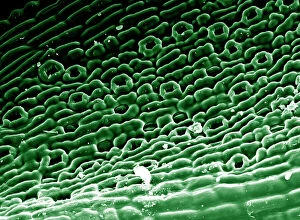
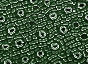
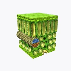
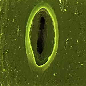
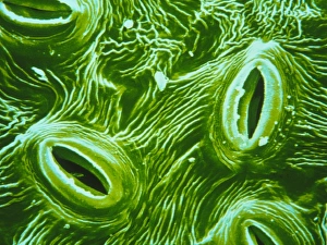
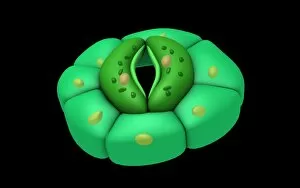
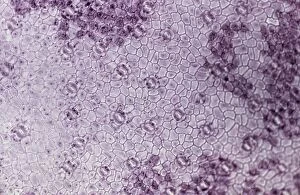
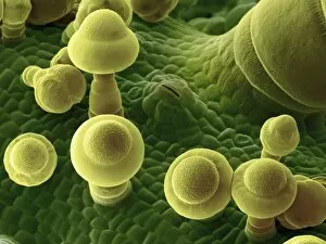
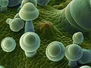
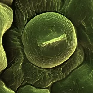
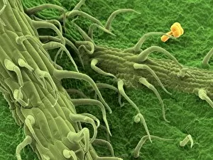
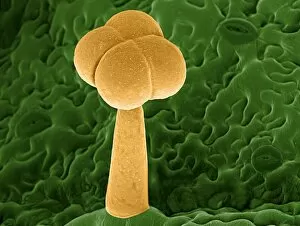
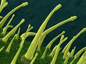
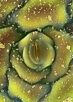
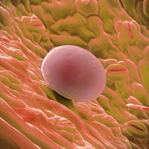
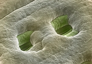
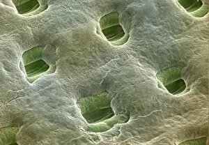
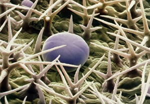
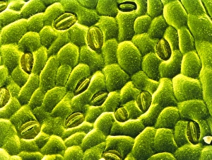
"Exploring the Fascinating World of Stomata: Leaf Pores Revealed Through SEM" In the microscopic realm, a hidden universe awaits our discovery - the intricate world of stomata. These tiny leaf pores, captured in stunning detail by scanning electron microscopy (SEM), offer us a glimpse into the remarkable structures that enable plants to breathe and regulate their vital processes. Picture No. 11675585 presents an enchanting view of English oak leaf pores under SEM. Delicately arranged like miniature mouths on the leaf's surface, these stomata serve as gatekeepers for gas exchange between plants and their environment. Moving on to Picture No. 11675628, we encounter French lavender leaf pore through SEM. Its elegant shape and structure showcase nature's precision at work, highlighting how each stoma is surrounded by specialized cells that control its opening and closing. Lemon grass takes center stage in another captivating image captured by SEM. With its elongated shape and distinct ridges, this stoma reveals itself as a testament to plant adaptation and survival strategies. Chives leaf stoma (SEM C016 / 8060) offers yet another intriguing perspective with its unique arrangement of guard cells surrounding the central opening. This intricate design allows for precise regulation of water loss while facilitating carbon dioxide uptake during photosynthesis. Coltsfoot leaf stomata (SEM C016 / 8052) present themselves as a mesmerizing pattern resembling celestial constellations scattered across the surface. Each individual pore represents an essential gateway for transpiration and gaseous exchange within this resilient plant species. Potato Leaf Stomata (SEM) showcases these minute structures embedded within potato leaves' epidermis – reminding us that even humble tubers possess complex mechanisms for respiration amidst their subterranean existence. Beyond gas exchange lies Geranium oil gland - an unexpected surprise amidst our exploration of stomatal wonders. This specialized structure secretes aromatic oils responsible for the plant's distinctive fragrance, adding a delightful twist to our journey.