
Strands Collection (page 4)
"Strands of Beauty: From the Northern Irish Coastline to Greek Sunsets" Embark on a journey along the picturesque strands that grace our world


All Professionally Made to Order for Quick Shipping
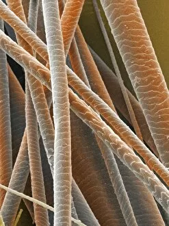
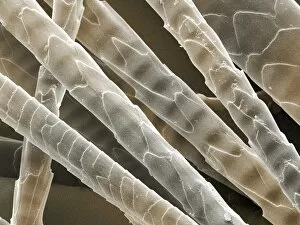
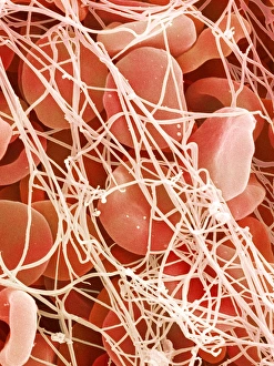
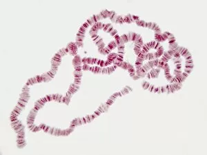
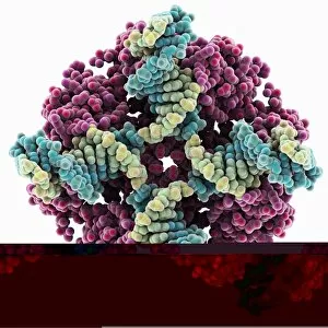
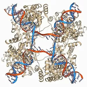
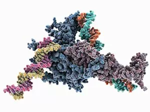
"Strands of Beauty: From the Northern Irish Coastline to Greek Sunsets" Embark on a journey along the picturesque strands that grace our world, weaving tales of natural wonders and scientific marvels. In Ballycastle, County Antrim, Northern Ireland, discover the breathtaking Northern Irish coastline with its wide sandy beaches. Here, nature's artistry is showcased in all its glory as golden sands meet crashing waves under an endless sky. Travel further south to Corfu Island in Greece and witness a mesmerizing sunset at Almyros Beach near Acharavi. As dusk descends upon this northern coast, vibrant hues paint the horizon while gentle waves kiss the shore - a sight that captures both heart and soul. Delve into the realm of science where strands take on a different form - DNA transcription. Molecular models reveal intricate patterns that hold life's secrets within their delicate structure. Like Trebarwith Strand in north Cornwall during high water at sunset, these molecular strands intertwine to create awe-inspiring beauty. From computer-generated models to artistic renditions, DNA molecules continue to captivate our imagination. Their elegant twists and turns symbolize life's complexity while reminding us of our interconnectedness with every living being. But not all they can made of molecules; some come in furry forms too. Picture a wet black Labrador Retriever bounding joyfully along the dog beach - his playful antics leaving trails behind him like footprints etched upon sand. Across continents we go next - Cannon Beach in Oregon offers panoramic views from Ecola State Park. Majestic rock formations rise from pristine shores as crashing waves carve new paths through time. Platja de Llenaire awaits us on Majorca's Balearic Islands where sandy beaches stretch out beneath azure skies. Tranquility reigns here as sun-seekers bask in warm rays while gentle tides caress their toes.