
Tetramer Collection
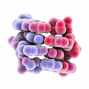
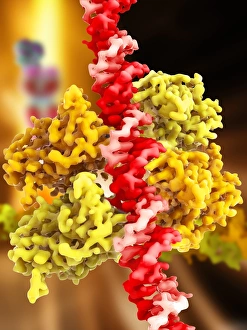
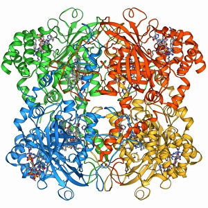
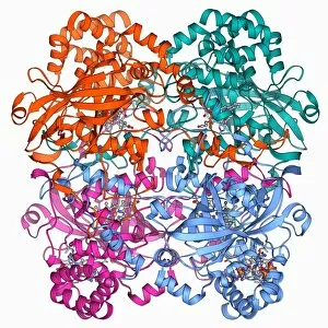
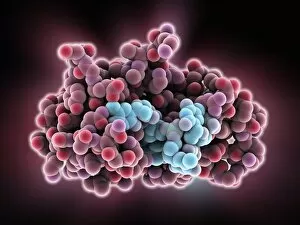
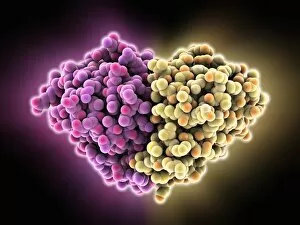
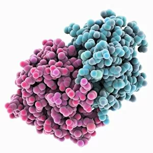
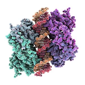
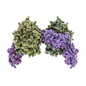
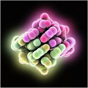
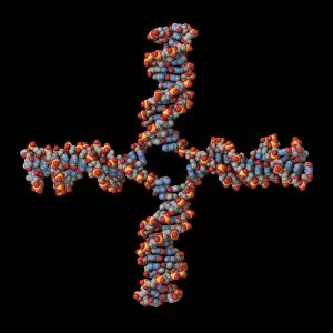
Caption: Exploring the Intricacies of Tetramers in Molecular Biology From left to right: Z-DNA tetramer molecule (C015 / 6557


All Professionally Made to Order for Quick Shipping
Caption: Exploring the Intricacies of Tetramers in Molecular Biology From left to right: Z-DNA tetramer molecule (C015 / 6557): Revealing the unique structure and properties of this fascinating DNA configuration. Tumour suppressor protein and DNA (C017 / 3647): Unraveling the intricate relationship between these crucial components in cancer prevention. Tumour suppressor protein molecular model: Gaining insights into the three-dimensional structure that plays a vital role in regulating cell growth and division. Catalase, molecular model (F006 / 9774): Understanding how this enzyme protects cells from oxidative stress by breaking down harmful hydrogen peroxide molecules. CAMP-dependent protein kinase molecule (F006 / 9728): Investigating the key player responsible for transmitting signals within cells, influencing various biological processes. 6 & Voltage-gated potassium channel (F006 / 9642 & F006 /9562): Delving into the mechanisms behind these channels' ability to regulate electrical impulses across cell membranes. 8 & Human catalase, molecular model (F006/9478) & Voltage-gated potassium channel (F006/9391): Examining human-specific structures with potential implications for disease research and drug development. 10 &11. Voltage-gated potassium channel (F006/9324) & (Human catalase, molecular model F006/9288 ): Shedding light on additional aspects of these critical proteins involved in maintaining cellular homeostasis. Carbamoylsarcosine amidase enzyme: Studying an essential enzyme involved in metabolizing carbamoyl compounds, potentially aiding therapeutic advancements. These captivating images showcase various facets of tetramers—molecules composed of four subunits—providing valuable insights into their structural intricacies and functional significance within biological systems.