
Thigh Bone Collection (page 3)
"The Thigh Bone: A Journey of Art, Replacement, and Resilience" Hip replacement meets artistry - a fusion of medical innovation and creative expression


All Professionally Made to Order for Quick Shipping
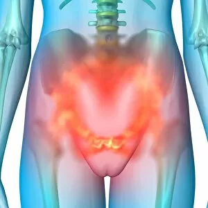
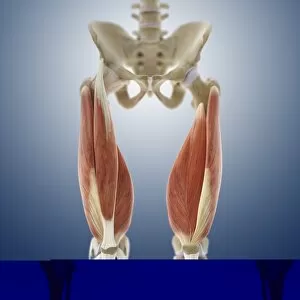
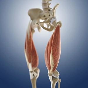
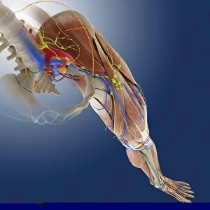
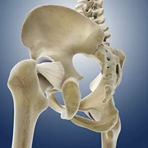
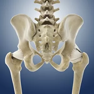
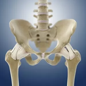
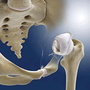
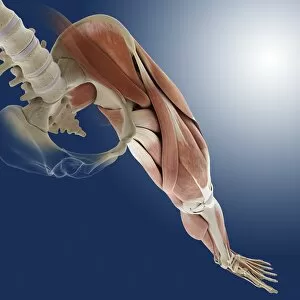
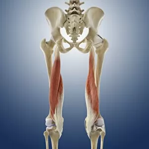
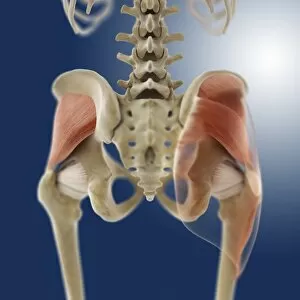
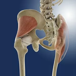
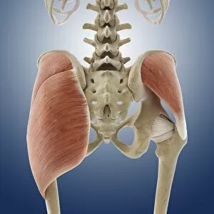
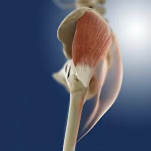
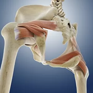
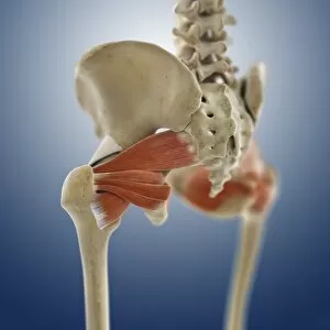
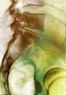
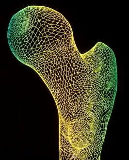
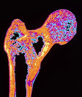
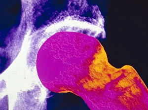
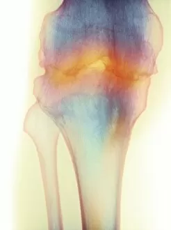
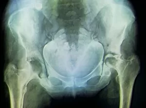
"The Thigh Bone: A Journey of Art, Replacement, and Resilience" Hip replacement meets artistry - a fusion of medical innovation and creative expression. Peering into the depths of healing - an X-ray reveals the success of total hip replacement surgery. From injury to inspiration - damaged knee ligament transformed into captivating artwork. Unveiling strength within - a healthy knee captured in its intricate beauty through an X-ray lens. Exploring the human frame - an enchanting engraving depicting the timeless elegance of our skeleton. Beneath the surface lies perfection - normal legs revealed through mesmerizing X-rays. Literary musings on anatomy's marvels - Henry Fielding's illustration brings Tom Jones to life alongside a femur bone engraving. Delving deeper for answers – CT scan captures the essence of a healthy knee in stunning detail (CT scan C018 / 0413). Celebrating power from behind – artistic portrayal showcases buttock muscles' strength and grace (artwork C013 / 4414). The foundation of our being unveiled – an exquisite illustration showcasing the majesty of human bones, including the mighty femur. Culinary exploration meets anatomy – using a kitchen knife to remove bone from chicken thigh highlights similarities between nature and ourselves. Osteoarthritis unraveled–an X-ray revealing F006/3744 showcases how it affects one's hip joint.