
Thirty Five Collection
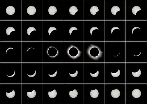
"Thirty Five: A Glimpse into the Past and Present" Step back in time to March 29, 2006, when a total solar eclipse mesmerized the world with its celestial beauty


All Professionally Made to Order for Quick Shipping
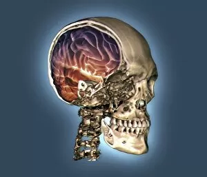
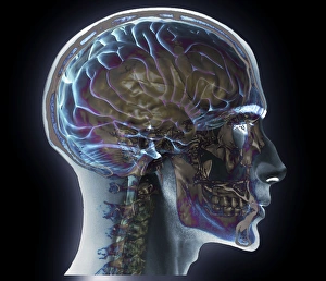
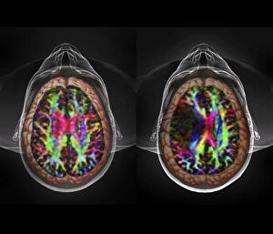
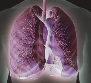
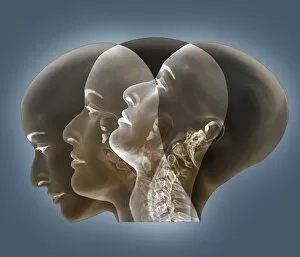
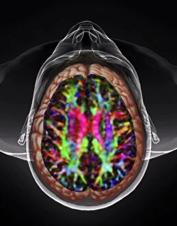
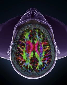
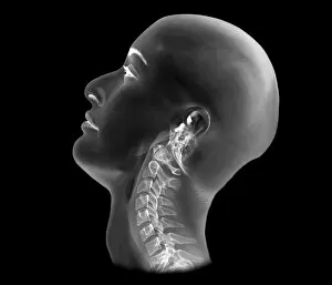
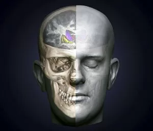
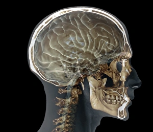
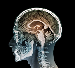
"Thirty Five: A Glimpse into the Past and Present" Step back in time to March 29, 2006, when a total solar eclipse mesmerized the world with its celestial beauty. As the moon perfectly aligned with the sun, darkness briefly engulfed our skies, leaving us in awe of nature's wonders. Transporting ourselves further back, we stumble upon a captivating Persian falconer tile panel from around 1910. Crafted by Pilkington's Tiles Group Plc using slip-coated earthenware, this masterpiece showcases the artistry and cultural richness of that era. Shifting gears to Hereford in 1904 brings us to an exhilarating event - the Automobile Club Small Car Trials. Witnessing these trials must have been a thrilling experience as small cars raced through challenging terrains, pushing boundaries and showcasing automotive innovation. Now let's delve into medical marvels that shed light on our own bodies. Behold a normal skull and brain captured through a remarkable 3D CT scan (C016/6333). This intricate imaging technique allows us to explore every nook and cranny of our complex neurological system. However, not all scans reveal such normalcy. The haunting presence of brain cancer is unveiled through DTI and 3D CT scans (C016/6414), reminding us of the fragility within even our most vital organ. Moving down to examine head and neck health becomes possible thanks to MRI and 3D CT scans (C016/6337) revealing what lies beneath our skin's surface. These advanced technologies provide invaluable insights for medical professionals striving to diagnose ailments accurately. A glimpse at X-rays unveils both normal flexed necks (C016/6426) and regular neck structures (C016/6429 & C016/6432). These images serve as reminders that even seemingly ordinary parts play crucial roles in maintaining overall well-being.