
Tract Collection (page 4)
"Exploring the Diverse Tracts of Knowledge: From Gallstones to Religious Engravings" Delve into history with the Brooklands Poster


All Professionally Made to Order for Quick Shipping
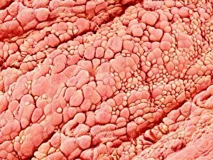
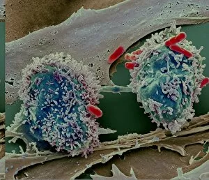
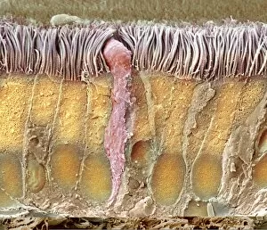
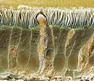
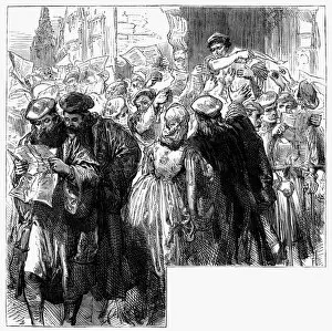
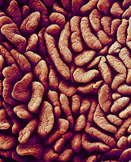
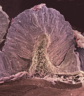
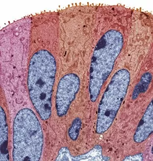
"Exploring the Diverse Tracts of Knowledge: From Gallstones to Religious Engravings" Delve into history with the Brooklands Poster, a captivating glimpse into motorsport's golden age. Uncover the secrets of our bodies through artwork like the intricate depiction of gallstones or a mesmerizing SEM image of the small intestine. Discover the therapeutic benefits of taking water at Bad Wildungen, Germany - an oasis for rejuvenation and healing. Dive deep into fish anatomy as you explore a historical model that unravels their fascinating inner workings. Unlock mysteries within our brains by examining white matter fibers through a cutting-edge DTI scan. Join renowned footballer Tim Cahill on his journey to success, as he leaves his own unique tract in sports history. Step back in time to Victorian England and witness Mary Jones with her Bible, symbolizing unwavering faith during an era defined by religious tracts. Chuckle at a comic postcard depicting a temperance speaker and their audience, shedding light on societal attitudes towards alcohol consumption in yesteryears. Reflect upon human vices through a Russian schematic illustration from 1850 showcasing the Seven Deadly Sins - reminding us of timeless moral tracts that shape society's conscience. Transport yourself to 1783 with "The Coalition-Dance, " capturing political satire through William Humphrey's hand-colored etching - offering insights into how tracts were used for political commentary. In this vast world filled with diverse tracts ranging from medical discoveries and historical artifacts to religious teachings and social commentaries, there is no shortage of knowledge waiting to be explored.