Fraud Blocker Skip to main content

Tumour Collection (page 2)

"Tumour: Unveiling the Battle Within" In a microscopic realm, T lymphocytes engage in an intense combat against cancer cells, as captured by SEM C001 / 1679

Background imageTumour Collection: Brain cancer, artwork F006 / 7483

Brain cancer, artwork F006 / 7483
Brain cancer, computer artwork

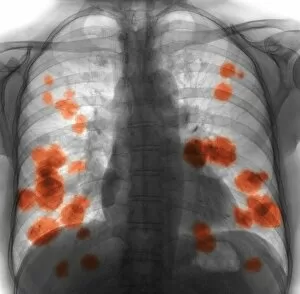
Background imageTumour Collection: Secondary lung cancer, X-ray F008 / 3498

Secondary lung cancer, X-ray F008 / 3498
Secondary lung cancer. Coloured X-ray of the chest of a 52 year old female patient with metastatic (secondary) lung cancer (red)

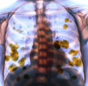
Background imageTumour Collection: Secondary lung cancer, X-ray F008 / 3496

Secondary lung cancer, X-ray F008 / 3496
Secondary lung cancer. Coloured X-ray of the chest of a 52 year old female patient with metastatic (secondary) lung cancer (yellow)

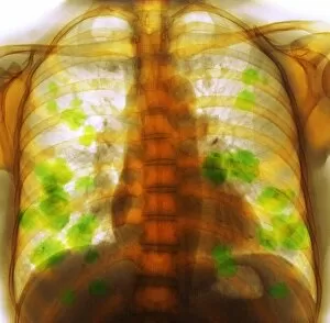
Background imageTumour Collection: Secondary lung cancer, X-ray F008 / 3495

Secondary lung cancer, X-ray F008 / 3495
Secondary lung cancer. Coloured X-ray of the chest of a 52 year old female patient with metastatic (secondary) lung cancer (green)

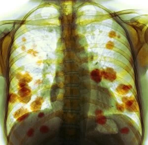
Background imageTumour Collection: Secondary lung cancer, X-ray F008 / 3497

Secondary lung cancer, X-ray F008 / 3497
Secondary lung cancer. Coloured X-ray of the chest of a 52 year old female patient with metastatic (secondary) lung cancer (orange)

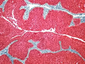
Background imageTumour Collection: Bladder cancer, light micrograph F005 / 6066

Bladder cancer, light micrograph F005 / 6066
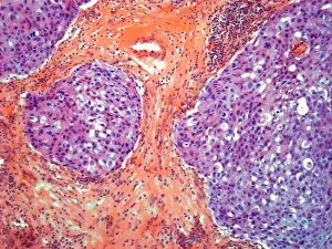
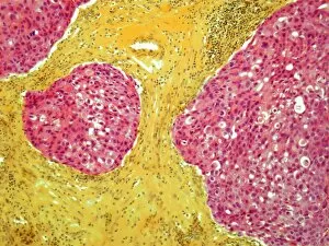
Bladder cancer. Light micrograph of a section through a transitional cell carcinoma (TCC, cancer) of the bladder wall. TCC is the most common form of bladder cancer

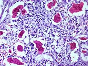
Background imageTumour Collection: Bone tumour, light micrograph F005 / 6096

Bone tumour, light micrograph F005 / 6096
Bone tumour. Light micrograph of a section through a giant cell tumour of the bone. This tumour is characterized by the presence of multinucleated giant cells (red)

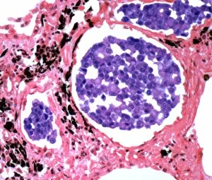
Background imageTumour Collection: Secondary lung cancer, light micrograph F005 / 6072

Secondary lung cancer, light micrograph F005 / 6072
Secondary lung cancer. Light micrograph of a section through a secondary carcinoma of the lung. This cancer has metastasised (spread) from a primary tumour of the breast
Background imageTumour Collection: Testicular cancer, light micrograph F005 / 6062

Testicular cancer, light micrograph F005 / 6062
Testicular cancer. Light micrograph of a section through a teratoma of the testis, a rare type of cancer. Teratomas are encapsulated malignant (cancerous)

Background imageTumour Collection: Testicular cancer, light micrograph F005 / 6063

Testicular cancer, light micrograph F005 / 6063
Testicular cancer. Light micrograph of a section through a teratoma of the testis, a rare type of cancer. Teratomas are encapsulated malignant (cancerous)

Background imageTumour Collection: Testicular cancer, light micrograph F005 / 6064

Testicular cancer, light micrograph F005 / 6064
Testicular cancer. Light micrograph of a section through a seminoma tumour of the testis. This is a germ cell tumour and one of the most treatable and curable cancers

Background imageTumour Collection: Prostate cancer, light micrograph F005 / 6093

Prostate cancer, light micrograph F005 / 6093
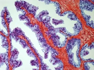
Prostate cancer. Light micrograph of a section through a carcinoma (cancer) of the prostate. Magnification: x200 when printed at 10 centimetres wide

Background imageTumour Collection: Thyroid cancer, light micrograph F005 / 6088

Thyroid cancer, light micrograph F005 / 6088
Thyroid cancer. Light micrograph of a section through a papillary adenocarcinoma (cancer) of the thyroid gland. Magnification: x200 when printed at 10 centimetres wide

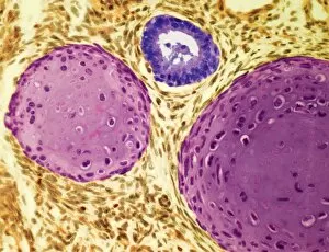
Background imageTumour Collection: Bone tumour, light micrograph F005 / 6097

Bone tumour, light micrograph F005 / 6097
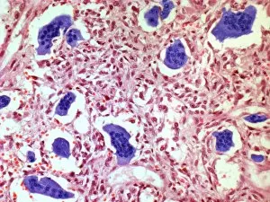
Bone tumour. Light micrograph of a section through a giant cell tumour of the bone. This tumour is characterized by the presence of multinucleated giant cells (purple)

Background imageTumour Collection: Breast cancer, light micrograph F005 / 6079

Breast cancer, light micrograph F005 / 6079
Breast cancer. Light micrograph of a section through a ductal carcinoma (cancer) of the breast. This is a cancer that arises from the lining of the milk ducts

Background imageTumour Collection: Bladder cancer, light micrograph F005 / 6065

Bladder cancer, light micrograph F005 / 6065
Bladder cancer. Light micrograph of a section through a transitional cell carcinoma (TCC, cancer) of the bladder wall. TCC is the most common form of bladder cancer

Background imageTumour Collection: Secondary liver cancer, light micrograph F005 / 6100

Secondary liver cancer, light micrograph F005 / 6100
Secondary liver cancer. Light micrograph of a section through liver tissue showing a secondary cancer that had metastasised (spread) from a primary small cell lung cancer

Background imageTumour Collection: Secondary heart cancer, light micrograph F005 / 6069

Secondary heart cancer, light micrograph F005 / 6069
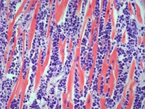
Secondary heart cancer. Light micrograph of a section through cardiac muscle showing a metastatic seminoma. This is a germ cell tumour that originated in the testis

Background imageTumour Collection: Breast cancer, artwork F005 / 0458

Breast cancer, artwork F005 / 0458
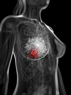
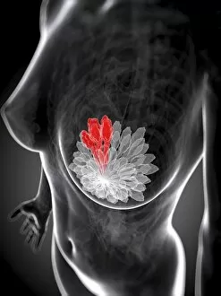
Breast cancer, computer artwork

Background imageTumour Collection: Breast cancer, artwork F005 / 0473

Breast cancer, artwork F005 / 0473
Breast cancer, computer artwork

Background imageTumour Collection: Bowel cancer, artwork F005 / 0525

Bowel cancer, artwork F005 / 0525
Bowel cancer, computer artwork

Background imageTumour Collection: Breast cancer, artwork F005 / 0466

Breast cancer, artwork F005 / 0466
Breast cancer, computer artwork

Background imageTumour Collection: Thyroid cancer, light micrograph F005 / 6087

Thyroid cancer, light micrograph F005 / 6087
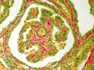
Thyroid cancer. Light micrograph of a section through a papillary adenocarcinoma (cancer) of the thyroid gland. Magnification: x200 when printed at 10 centimetres wide

Background imageTumour Collection: Breast cancer, light micrograph F005 / 6080

Breast cancer, light micrograph F005 / 6080
Breast cancer. Light micrograph of a section through a ductal carcinoma (cancer) of the breast. This is a cancer that arises from the lining of the milk ducts

Background imageTumour Collection: Kidney cancer, light micrograph F005 / 6071

Kidney cancer, light micrograph F005 / 6071
Kidney cancer. Light micrograph of a section through a Wilms tumour of the kidney. This solid tumour generally affects children, with most cases occurring between 3 and 4 years of age

Background imageTumour Collection: Uterine cancer, light micrograph F005 / 6074

Uterine cancer, light micrograph F005 / 6074
Uterine cancer. Light micrograph of a section through an adenocarcinoma (cancer) of the endometrium (womb lining). Magnification: x300 when printed at 10 centimetres

Background imageTumour Collection: Uterine cancer, light micrograph F005 / 6073

Uterine cancer, light micrograph F005 / 6073
Uterine cancer. Light micrograph of a section through an adenocarcinoma (cancer) of the endometrium (womb lining). Magnification: x300 when printed at 10 centimetres

Background imageTumour Collection: Breast cancer, artwork F005 / 0459

Breast cancer, artwork F005 / 0459
Breast cancer, computer artwork

Background imageTumour Collection: Breast cancer, artwork F005 / 0471

Breast cancer, artwork F005 / 0471
Breast cancer, computer artwork

Background imageTumour Collection: Breast cancer, artwork F005 / 0463

Breast cancer, artwork F005 / 0463
Breast cancer, computer artwork

Background imageTumour Collection: Breast cancer, artwork F005 / 0464

Breast cancer, artwork F005 / 0464
Breast cancer, computer artwork

Background imageTumour Collection: Breast cancer, artwork F005 / 0461

Breast cancer, artwork F005 / 0461
Breast cancer, computer artwork

Background imageTumour Collection: Breast cancer, artwork F005 / 0470

Breast cancer, artwork F005 / 0470
Breast cancer, computer artwork

Background imageTumour Collection: Breast cancer, artwork F005 / 0462

Breast cancer, artwork F005 / 0462
Breast cancer, computer artwork

Background imageTumour Collection: Breast cancer, artwork F005 / 0472

Breast cancer, artwork F005 / 0472
Breast cancer, computer artwork

Background imageTumour Collection: Breast cancer, artwork F005 / 0469

Breast cancer, artwork F005 / 0469
Breast cancer, computer artwork

Background imageTumour Collection: Breast cancer, artwork F005 / 0457

Breast cancer, artwork F005 / 0457
Breast cancer, computer artwork

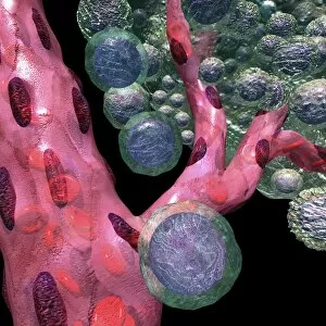
Background imageTumour Collection: Blood vessel formation, artwork C016 / 9842

Blood vessel formation, artwork C016 / 9842
Blood vessel formation. Artwork showing malignant (cancerous) tumour cells (green) promoting the formation of new blood vessels (pink), a process known as angiogenesis

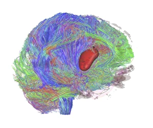
Background imageTumour Collection: Brain tumour, DTI MRI scan C017 / 7057

Brain tumour, DTI MRI scan C017 / 7057
Brain tumour. 3D diffusion tensor imaging (DTI) magnetic resonance imaging (MRI) scan of nerve pathways (coloured) in a brain with a tumour (red, centre right)

Background imageTumour Collection: Uterine cancer, artwork F006 / 2550

Uterine cancer, artwork F006 / 2550
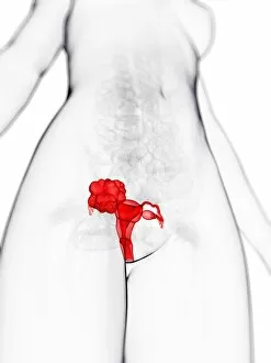
Uterine cancer, computer artwork

Background imageTumour Collection: Uterine cancer, artwork F006 / 2551

Uterine cancer, artwork F006 / 2551
Uterine cancer, computer artwork

Background imageTumour Collection: Healthy thyroid, artwork F006 / 2526

Healthy thyroid, artwork F006 / 2526
Healthy thyroid, computer artwork

Background imageTumour Collection: Thyroid cancer, artwork F006 / 2527

Thyroid cancer, artwork F006 / 2527
Thyroid cancer, computer artwork

Background imageTumour Collection: Stomach cancer, artwork F006 / 2513

Stomach cancer, artwork F006 / 2513
Stomach cancer, computer artwork

Background imageTumour Collection: Spleen cancer, artwork F006 / 2505

Spleen cancer, artwork F006 / 2505
Spleen cancer, computer artwork

Background imageTumour Collection: Breast cancer, artwork F006 / 2386

Breast cancer, artwork F006 / 2386
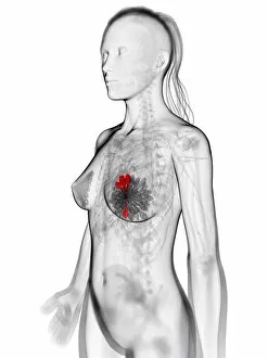
Breast cancer, computer artwork

Background imageTumour Collection: Lung cancer, artwork F006 / 2376

Lung cancer, artwork F006 / 2376
Lung cancer, computer artwork

Background imageTumour Collection: Lung cancer, artwork F006 / 2375

Lung cancer, artwork F006 / 2375
Lung cancer, computer artwork


All Professionally Made to Order for Quick Shipping

"Tumour: Unveiling the Battle Within" In a microscopic realm, T lymphocytes engage in an intense combat against cancer cells, as captured by SEM C001 / 1679. These brave warriors of our immune system tirelessly fight to protect us from the clutches of disease. Moving to a different battleground, ovarian cancer reveals its intricate nature under the lens of a light micrograph (C015 / 7103). This visual exploration sheds light on the complexity and urgency surrounding this formidable adversary. Amidst these scientific endeavors, nanorobots emerge as potential allies in our war against cancer. Witness their futuristic assault on malignant cells, offering hope for a brighter future where tumors are eradicated (Nanorobot attacking cancer). Beyond the realm of science lies another facet of tumours - congenital eye anomalies. Klinische Darstellungen der provides insight into these mysterious conditions that affect vision and challenge medical professionals' understanding. Exploring further, we encounter "Kyste sereux cloisonne du cou" and "Lipome de la tete, " captivating colour lithographs that depict unique manifestations of tumours in unexpected places. These artistic representations remind us that even within chaos lies beauty waiting to be discovered. Delving into history's archives brings forth Joanna Southcott, an extraordinary fanatic whose beliefs stirred controversy during her time. Like a metaphorical tumour challenging societal norms, she left an indelible mark on history (Joanna Southcott: An Extraordinary Fanatic, 1822). Artistic expressions also capture the essence of tumours; J-L David's masterpiece Chevignard portrays emotions intertwined with physical affliction caused by these growths. Through art's lens emerges empathy for those affected by such ailments (J-L David / Chevignard). Nature too holds its own share of peculiarities – behold Catasetum callosum with its tumor-lipped or callous appearance.