Fraud Blocker Skip to main content

Tumour Collection (page 6)

"Tumour: Unveiling the Battle Within" In a microscopic realm, T lymphocytes engage in an intense combat against cancer cells, as captured by SEM C001 / 1679

Background imageTumour Collection: Dermoid ovarian cyst C015 / 6056

Dermoid ovarian cyst C015 / 6056
Dermoid ovarian cyst. Light micrograph of a section through an ovary with a dermoid, or teratoma, ovarian cyst. This is a benign (non-cancerous) cyst that arises from germ (sex) cells

Background imageTumour Collection: Mesothelioma lung cancer, CT scan

Mesothelioma lung cancer, CT scan
Mesothelioma lung cancer. Computed tomography (CT) scan of the lungs of a 69 year old patient with mesothelioma, a type of lung cancer

Background imageTumour Collection: Lung cancer, CT and PET scans

Lung cancer, CT and PET scans
Lung cancer. Combined computed tomography (CT) and positron emission tomography (PET) scans of the lungs of an 84 year old woman with a malignant (cancerous) tumour (bright patch)

Background imageTumour Collection: Intestinal tumour, artwork C015 / 6451

Intestinal tumour, artwork C015 / 6451
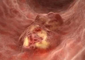
Intestinal tumour. Computer artwork of a tumour on the internal wall of an intestine

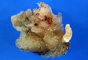
Background imageTumour Collection: Spleen cancer, gross specimen C015 / 6421

Spleen cancer, gross specimen C015 / 6421
Spleen cancer. Gross specimen of a spleen showing a large lymphosarcoma, or non-Hodgkins lymphoma, a cancer of lymphoid tissue. The lymphatic system is a major part of the immune system
Background imageTumour Collection: Uterine fibroid C015 / 6416

Uterine fibroid C015 / 6416
Uterine fibroid. Gross specimen of a sectioned uterine fibroid. A fibroid is a fibrous benign tumour originating from muscular tissue

Background imageTumour Collection: Uterine fibroid, light micrograph C015 / 6413

Uterine fibroid, light micrograph C015 / 6413
Uterine fibroid. Light micrograph of a section through a uterine fibroid. A fibroid is a fibrous benign tumour originating from muscular tissue

Background imageTumour Collection: Uterine fibroid C015 / 6415

Uterine fibroid C015 / 6415
Uterine fibroid. Gross specimen of a sectioned uterine fibroid. A fibroid is a fibrous benign tumour originating from muscular tissue

Background imageTumour Collection: Uterine fibroid, light micrograph C015 / 6411

Uterine fibroid, light micrograph C015 / 6411
Uterine fibroid. Light micrograph of a section through a uterine fibroid (centre). A fibroid is a fibrous benign tumour originating from muscular tissue

Background imageTumour Collection: Ovarian fibroid C015 / 6414

Ovarian fibroid C015 / 6414
Ovarian fibroid. Gross specimen of an ovarian fibroid. A fibroid is a fibrous benign tumour originating from muscular tissue

Background imageTumour Collection: Uterine fibroid, light micrograph C015 / 6412

Uterine fibroid, light micrograph C015 / 6412
Uterine fibroid. Light micrograph of a section through a uterine fibroid (oval). A fibroid is a fibrous benign tumour originating from muscular tissue

Background imageTumour Collection: Uterine fibroid C015 / 6406

Uterine fibroid C015 / 6406
Uterine fibroid. Gross specimen of a uterine fibroid. A fibroid is a fibrous benign tumour originating from muscular tissue

Background imageTumour Collection: Dermoid ovarian cyst C015 / 6402

Dermoid ovarian cyst C015 / 6402
Dermoid ovarian cyst. Light micrograph of a section through an ovary with a dermoid, or teratoma, ovarian cyst. This is a benign (non-cancerous) cyst that arises from germ (sex) cells

Background imageTumour Collection: Bladder mass, ultrasound scan C017 / 7796

Bladder mass, ultrasound scan C017 / 7796
Bladder mass. Ultrasound scan showing a large mass (centre) in the bladder of an 84 year old male patient. This could be a malignant (cancerous) tumour

Background imageTumour Collection: Bladder mass, Doppler ultrasound C017 / 7795

Bladder mass, Doppler ultrasound C017 / 7795
Bladder mass. Doppler ultrasound scan showing a large mass (black) with increased blood flow in the bladder of an 84 year old male patient. This could be a malignant (cancerous) tumour

Background imageTumour Collection: Mesothelioma lung cancer, X-ray C017 / 7776

Mesothelioma lung cancer, X-ray C017 / 7776
Mesothelioma lung cancer, X-ray of the chest of a 84 year old male patient with advanced mesothelioma, a type of lung cancer

Background imageTumour Collection: Angiosarcoma of the spleen C015 / 6398

Angiosarcoma of the spleen C015 / 6398
Angiosarcoma. Gross specimen of a sectioned spleen affected by angiosarcoma. Malignant (cancerous) tumours (yellow) can be seen at bottom centre

Background imageTumour Collection: Secondary pancreatic cancer, ultrasound C017 / 7765

Secondary pancreatic cancer, ultrasound C017 / 7765
Secondary pancreatic cancer. Ultrasound scan of a 63 year old male patients pancreas showing metastatic (secondary) tumours. The cancer has metastasised (spread) from a primary cancer

Background imageTumour Collection: Secondary thyroid cancer C015 / 6170

Secondary thyroid cancer C015 / 6170
Secondary thyroid cancer. Light micrograph of a section through a tumour in a lymph node due to a secondary thyroid cancer

Background imageTumour Collection: Alveolar soft part sarcoma, TEM C016 / 5826

Alveolar soft part sarcoma, TEM C016 / 5826
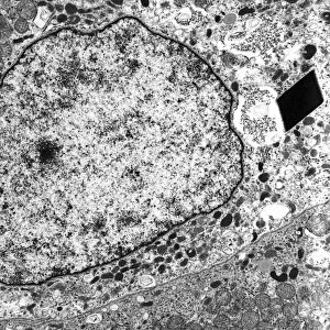
Alveolar soft part sarcoma. Transmission electron micrograph (TEM) of a section through an alveolar soft part sarcoma (ASPS) showing a characteristic rhomboid crystal (black, centre)

Background imageTumour Collection: Alveolar soft part sarcoma, TEM C016 / 5816

Alveolar soft part sarcoma, TEM C016 / 5816
Alveolar soft part sarcoma. Transmission electron micrograph (TEM) of a section through an alveolar soft part sarcoma (ASPS) showing a characteristic rhomboid crystal (black)

Background imageTumour Collection: Brain cancer, MRI scan C016 / 4438

Brain cancer, MRI scan C016 / 4438
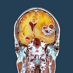
Brain cancer. Coloured coronal magnetic resonance imaging (MRI) scan through the head of a 48 year old male patient with a glioblastoma (red)

Background imageTumour Collection: Brain cancer, MRI scan C016 / 4436

Brain cancer, MRI scan C016 / 4436
Brain cancer. Coloured coronal magnetic resonance imaging (MRI) scan through the head of a 48 year old male patient with a glioblastoma (centre right)

Background imageTumour Collection: Brain cancer, MRI scan C016 / 4437

Brain cancer, MRI scan C016 / 4437
Brain cancer. Coloured coronal magnetic resonance imaging (MRI) scan through the head of a 48 year old male patient with a glioblastoma (orange)

Background imageTumour Collection: Lung cancer, X-ray C016 / 4435

Lung cancer, X-ray C016 / 4435
Lung cancer. Coloured chest X-ray of a 64 year old male patient with lung cancer. The tumour is seen as the red mass in the right lung (bright area at left)

Background imageTumour Collection: Lung cancer, X-ray C016 / 4432

Lung cancer, X-ray C016 / 4432
Lung cancer. Coloured chest X-ray of a 64 year old male patient with lung cancer. The tumour is seen as the orange mass in the right lung (dark area at left)

Background imageTumour Collection: Lung cancer, X-ray C016 / 4433

Lung cancer, X-ray C016 / 4433
Lung cancer. Coloured chest X-ray of a 64 year old male patient with lung cancer. The tumour is seen as the orange mass in the right lung (bright area at left)

Background imageTumour Collection: Human tumour protein p73 C016 / 2830

Human tumour protein p73 C016 / 2830
Human tumour protein p73. Molecular model of the human tumour protein p73, encoded by the TP73 gene. Like the similar p53, is is a transcription factor of a type known as a tumour suppressor gene

Background imageTumour Collection: Human tumour protein p73 C016 / 2831

Human tumour protein p73 C016 / 2831
Human tumour protein p73. Molecular model of the human tumour protein p73, encoded by the TP73 gene. Like the similar p53, is is a transcription factor of a type known as a tumour suppressor gene

Background imageTumour Collection: Human tumour protein p63 C016 / 2681

Human tumour protein p63 C016 / 2681
Human tumour protein p63. Molecular model of the tetramerization domain of the human tumour protein p63, encoded by the TP63 gene

Background imageTumour Collection: Human tumour protein p63 C016 / 2680

Human tumour protein p63 C016 / 2680
Human tumour protein p63. Molecular model of the tetramerization domain of the human tumour protein p63, encoded by the TP63 gene

Background imageTumour Collection: Bowel disease, artwork C013 / 4611

Bowel disease, artwork C013 / 4611
Bowel disease. Computer artwork of an abnormal growth and inflammation in the lining of the caecum in the large intestine

Background imageTumour Collection: Bowel disease, artwork C013 / 4610

Bowel disease, artwork C013 / 4610
Bowel disease. Computer artwork of an abnormal growth and inflammation in the lining of the caecum in the large intestine

Background imageTumour Collection: Bowel disease, artwork C013 / 4609

Bowel disease, artwork C013 / 4609
Bowel disease. Computer artwork of an abnormal growth and inflammation in the lining of the caecum in the large intestine

Background imageTumour Collection: Agnews surgical clinic, 1889

Agnews surgical clinic, 1889
Agnews surgical clinic. At left, holding a scalpel, is US surgeon David Hayes Agnew (1818-1892), supervising a partial breast removal operation (mastectomy)

Background imageTumour Collection: T lymphocytes and cancer cells, SEM

T lymphocytes and cancer cells, SEM
T lymphocytes and cancer cells. Coloured scanning electron micrograph (SEM) of T lymphocyte cells (purple) attached to cancer cells

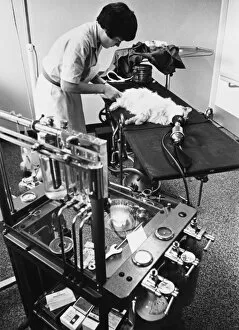
Background imageTumour Collection: Cat on Operating Table

Cat on Operating Table
A young veterinary nurse, Elizabeth Greaves, 17, bandages the leg of an unconscious cat, which has just had an operation to remove a tumour from its leg Date: late 1960s

Background imageTumour Collection: Great Tit with tumor growth on throat/chin

Great Tit with tumor growth on throat/chin

Background imageTumour Collection: Brain nausea reflex

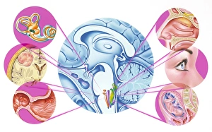
Brain nausea reflex. Artwork of the location in the brain of the area responsible for the various nausea reflexes, and some of the causes

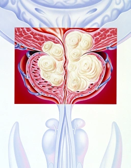
Background imageTumour Collection: Illustration of a benign prostatic hyperplasia

Illustration of a benign prostatic hyperplasia

Background imageTumour Collection: Uterine fibroid, MRI scan

Uterine fibroid, MRI scan

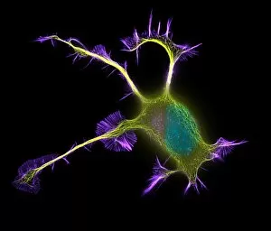
Background imageTumour Collection: Nerve cell growth

Nerve cell growth. Fluorescent light micrograph of a PC12 cell following stimulation by nerve growth factor. The cell body contains the nucleus (green)

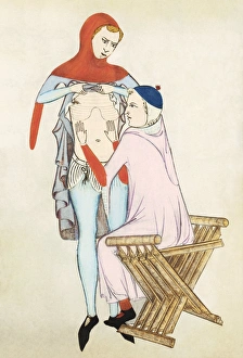
Background imageTumour Collection: Patient examination, 14th century artwork

Patient examination, 14th century artwork
Patient examination. 14th century artwork of a doctor examining a patient by palpation of the abdomen. Artwork from Anathomia (1345), by the Italian anatomist Guido da Vigevano

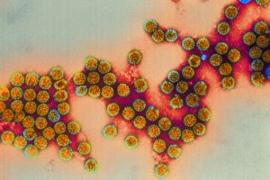
Background imageTumour Collection: Polyoma viruses

Polyoma viruses. Coloured transmission electron micrograph (TEM) of polyoma viruses. These simian (monkey) viruses (SV40) are a type of papovavirus

Background imageTumour Collection: Aphasia artwork

Aphasia artwork
Aphasia. Conceptual computer artwork of a blindfolded and gagged man representing the disability of aphasia. Aphasia is an impairment of language affecting the production or comprehension of speech

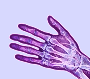
Background imageTumour Collection: Osteoid osteoma, X-ray

Osteoid osteoma, X-ray
Osteoid osteoma. Coloured X-ray of an osteoid osteoma (white, centre left) in the second phalange bone of the middle finger

Background imageTumour Collection: Gross specimen: atrial myxoma, benign heart tumour

Gross specimen: atrial myxoma, benign heart tumour
Atrial myxoma. Gross specimen of an atrial myxoma, a benign, jelly-like tumour of the connective tissue found in the heart

Background imageTumour Collection: Osteoid osteoma, light micrograph

Osteoid osteoma, light micrograph
Osteoid osteoma. Light micrograph of a section through an osteoid osteoma, a rare benign (non-cancerous) tumour of bone tissue that is very painful


All Professionally Made to Order for Quick Shipping

"Tumour: Unveiling the Battle Within" In a microscopic realm, T lymphocytes engage in an intense combat against cancer cells, as captured by SEM C001 / 1679. These brave warriors of our immune system tirelessly fight to protect us from the clutches of disease. Moving to a different battleground, ovarian cancer reveals its intricate nature under the lens of a light micrograph (C015 / 7103). This visual exploration sheds light on the complexity and urgency surrounding this formidable adversary. Amidst these scientific endeavors, nanorobots emerge as potential allies in our war against cancer. Witness their futuristic assault on malignant cells, offering hope for a brighter future where tumors are eradicated (Nanorobot attacking cancer). Beyond the realm of science lies another facet of tumours - congenital eye anomalies. Klinische Darstellungen der provides insight into these mysterious conditions that affect vision and challenge medical professionals' understanding. Exploring further, we encounter "Kyste sereux cloisonne du cou" and "Lipome de la tete, " captivating colour lithographs that depict unique manifestations of tumours in unexpected places. These artistic representations remind us that even within chaos lies beauty waiting to be discovered. Delving into history's archives brings forth Joanna Southcott, an extraordinary fanatic whose beliefs stirred controversy during her time. Like a metaphorical tumour challenging societal norms, she left an indelible mark on history (Joanna Southcott: An Extraordinary Fanatic, 1822). Artistic expressions also capture the essence of tumours; J-L David's masterpiece Chevignard portrays emotions intertwined with physical affliction caused by these growths. Through art's lens emerges empathy for those affected by such ailments (J-L David / Chevignard). Nature too holds its own share of peculiarities – behold Catasetum callosum with its tumor-lipped or callous appearance.