
Under Side Collection (page 4)
"Exploring the Hidden Beauty: From George Mutch's Goal to Underneath a Crowned Hairstreak Butterfly, Red Squirrel, and More


All Professionally Made to Order for Quick Shipping
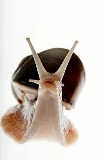
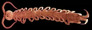
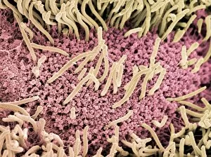
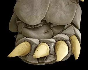
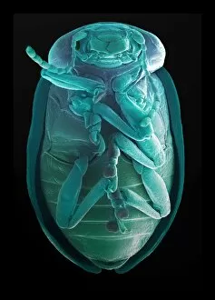
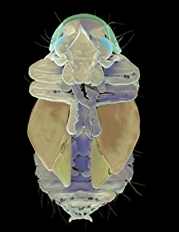
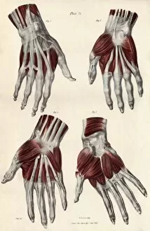
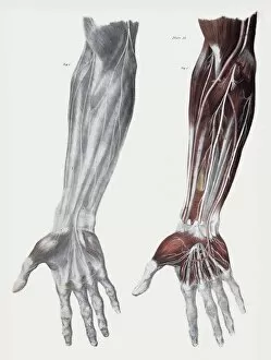
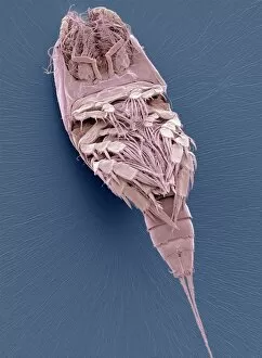
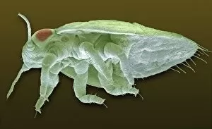
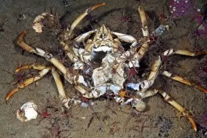
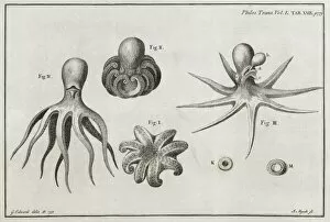
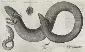
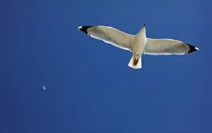
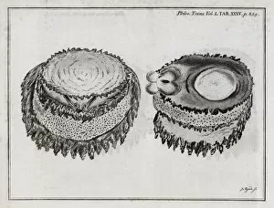
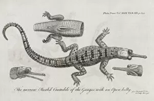
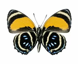
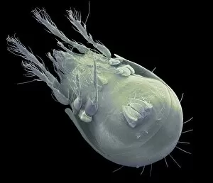
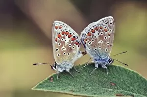
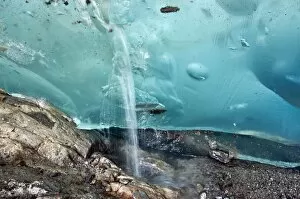
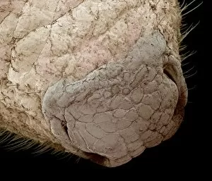
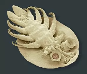
"Exploring the Hidden Beauty: From George Mutch's Goal to Underneath a Crowned Hairstreak Butterfly, Red Squirrel, and More. " Diving into the under side of captivating moments, we witness George Mutch's triumphant goal for Preston North End at the FA Cup Final. But let us not forget that beneath our feet lies a mesmerizing world filled with wonders. Delicate and elusive, a crowned hairstreak butterfly reveals its vibrant colors as it gracefully flutters through the air. Meanwhile, a red squirrel perches on a branch, showcasing its playful nature amidst nature's backdrop. Transporting ourselves back in time through an Air Mail Poster, we uncover Hensons Aerial Steam Carriage - an invention ahead of its time. Just like this remarkable contraption, there is so much more beneath the surface waiting to be discovered. Inquisitive jackdaws peck away at hidden treasures while giant manta rays glide effortlessly through ocean depths. Their majestic presence reminds us of the vastness below our everyday lives. Venturing further into this mysterious realm unveils intricate skull anatomy and delicate sulphur butterflies (Phoebis sp. ) dancing among blossoms. Moon jellyfish pulsate gently in ethereal beauty as if painting strokes across their underwater canvas. Finally, our journey concludes with an awe-inspiring sight overhead - a giant manta ray soaring gracefully against Mexico's Revillagigedo Islands' backdrop. This breathtaking encounter leaves us humbled by Mother Nature's grandeur from every angle. As we embrace these glimpses into the under side of life itself, may we remember that beyond what meets the eye lies endless marvels awaiting exploration – reminding us that true beauty often resides where least expected.