Fraud Blocker Skip to main content

Urinary System Collection (page 8)

The urinary system is a complex network of organs that play a vital role in maintaining the body's overall health and balance

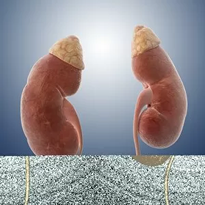
Background imageUrinary System Collection: Healthy adrenal glands, artwork

Healthy adrenal glands, artwork
Healthy adrenal glands, computer artwork

Background imageUrinary System Collection: Male urinary system, artwork F006 / 8468

Male urinary system, artwork F006 / 8468
Male urinary system, computer artwork

Background imageUrinary System Collection: Male urinary system, artwork F006 / 8422

Male urinary system, artwork F006 / 8422
Male urinary system, computer artwork

Background imageUrinary System Collection: Male urinary system, artwork F006 / 8403

Male urinary system, artwork F006 / 8403
Male urinary system, computer artwork

Background imageUrinary System Collection: Male urinary system, artwork F006 / 8392

Male urinary system, artwork F006 / 8392
Male urinary system, computer artwork

Background imageUrinary System Collection: Male urinary system, artwork F006 / 8391

Male urinary system, artwork F006 / 8391
Male urinary system, computer artwork

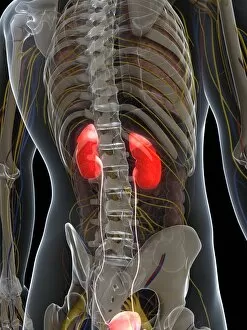
Background imageUrinary System Collection: Healthy kidneys, artwork F006 / 7613

Healthy kidneys, artwork F006 / 7613
Healthy kidneys, computer artwork

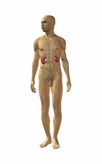
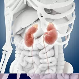
Background imageUrinary System Collection: Kidneys, anatomical artwork

Kidneys, anatomical artwork
Kidneys. Anatomical computer artwork of a mans body, showing the position of the kidneys (red, centre left and right) and adrenal glands (beige)

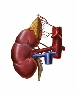
Background imageUrinary System Collection: Renal blood supply, anatomical artwork

Renal blood supply, anatomical artwork
Renal blood supply. Anatomical computer artwork of a human kidney (left) and adrenal gland (beige, upper centre), showing their main blood supply

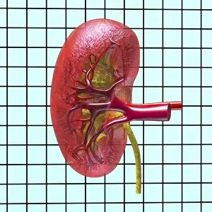
Background imageUrinary System Collection: Kidney anatomy, artwork C016 / 8945

Kidney anatomy, artwork C016 / 8945
Kidney anatomy, artwork. The kidneys filter waste from the blood and excrete it as urine. The kidney consists of an outer cortex and an inner medulla

Background imageUrinary System Collection: Kidney stones, artwork C016 / 7531

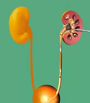
Kidney stones, artwork C016 / 7531
Kidney stones. Computer artwork showing a section through a human kidney containing kidney stones (yellowish, right). The ureter (tube)

Background imageUrinary System Collection: Incontinence surgery, artwork C016 / 6843

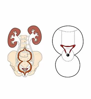
Incontinence surgery, artwork C016 / 6843
Incontinence surgery. Artwork of the sacral region of a womans pelvis showing a schematic diagram for bladder and sacral surgery to treat incontinence

Background imageUrinary System Collection: Inflamed kidney

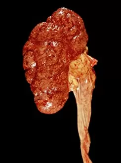
Inflamed kidney
Inflamed and swollen kidney, gross specimen. This is a case of chronic nephritis, an inflammation of the kidney. Nephritis is often caused by a bacterial infection

Background imageUrinary System Collection: Bladder adhesions

Bladder adhesions. Gross specimen of a urinary bladder with adhesions (string-like) across its inner surface. Adhesions are growths of fibrous (scar)

Background imageUrinary System Collection: Voiding cystourethrogram, X-rays C018 / 0577

Voiding cystourethrogram, X-rays C018 / 0577
Voiding cystourethrogram. X-ray of a 54 year old male patient undergoing a voiding cystourethrogram. This procedure involves liquid containing a radio-opaque dye being placed in the bladder via a

Background imageUrinary System Collection: Voiding cystourethrogram, X-rays C018 / 0578

Voiding cystourethrogram, X-rays C018 / 0578
Voiding cystourethrogram. Coloured X-ray of a 54 year old male patient undergoing a voiding cystourethrogram. This procedure involves liquid containing a radio-opaque dye being placed in the bladder

Background imageUrinary System Collection: Voiding cystourethrogram, X-rays C018 / 0579

Voiding cystourethrogram, X-rays C018 / 0579
Voiding cystourethrogram. Coloured X-ray of a 54 year old male patient undergoing a voiding cystourethrogram. This procedure involves liquid containing a radio-opaque dye being placed in the bladder

Background imageUrinary System Collection: Voiding cystourethrogram, X-rays C018 / 0465

Voiding cystourethrogram, X-rays C018 / 0465
Voiding cystourethrogram. Three coloured X-rays showing a 65 year old patient undergoing a voiding cystourethrogram. This procedure involves liquid containing a radio-opaque dye being placed in

Background imageUrinary System Collection: Nephron structure, artwork C016 / 9867

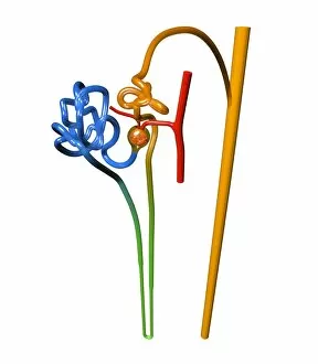
Nephron structure, artwork C016 / 9867
Nephron structure. Artwork showing the structure of a nephron, the functional filtering unit of a kidney. The function of the kidney is to filter waste products such as urea from the blood

Background imageUrinary System Collection: Nephron structure, artwork C016 / 9868

Nephron structure, artwork C016 / 9868
Nephron structure. Artwork showing the structure of a nephron, the functional filtering unit of a kidney. The function of the kidney is to filter waste products such as urea from the blood

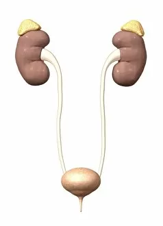
Background imageUrinary System Collection: Healthy kidneys, artwork C014 / 0397

Healthy kidneys, artwork C014 / 0397
Healthy kidneys, computer artwork. The kidneys filter waste products from the blood. The waste products are transported as urine to the bladder (not seen) via the ureters (tubes)

Background imageUrinary System Collection: Healthy kidneys, artwork C014 / 0398

Healthy kidneys, artwork C014 / 0398
Healthy kidneys, computer artwork. The kidneys filter waste products from the blood. The waste products are transported as urine to the bladder (not seen) via the ureters (tubes)

Background imageUrinary System Collection: Human kidneys, historical model

Human kidneys, historical model
Human kidneys. 1950s anatomical model of the human kidneys (red) with the ureters (tubes, upper left) attached, and a section through one kidney showing the internal structure

Background imageUrinary System Collection: Kidney stones, artwork

Kidney stones, artwork
Kidney stones. Computer artwork showing a section through a human kidney containing kidney stones (whiteish). The ureter (lower left)

Background imageUrinary System Collection: Bladder stones, artwork

Bladder stones, artwork
Bladder stones. Computer artwork showing a section through a human bladder containing bladder stones (whiteish, centre). The urethra can be seen (vertical, lower centre)

Background imageUrinary System Collection: Kidney anatomy, artwork C016 / 8663

Kidney anatomy, artwork C016 / 8663
Kidney anatomy, artwork. The kidneys filter waste from the blood and excrete it as urine. The kidney consists of an outer cortex and an inner medulla

Background imageUrinary System Collection: Kidney anatomy, artwork C016 / 8661

Kidney anatomy, artwork C016 / 8661
Kidney anatomy, artwork. The kidneys filter waste from the blood and excrete it as urine. The kidney consists of an outer cortex and an inner medulla

Background imageUrinary System Collection: Kidney anatomy, artwork C016 / 8662

Kidney anatomy, artwork C016 / 8662
Kidney anatomy, artwork. The kidneys filter waste from the blood and excrete it as urine. The kidney consists of an outer cortex and an inner medulla

Background imageUrinary System Collection: Kidney anatomy, artwork C016 / 8660

Kidney anatomy, artwork C016 / 8660
Kidney anatomy, artwork. The kidneys filter waste from the blood and excrete it as urine. The kidney consists of an outer cortex and an inner medulla

Background imageUrinary System Collection: Kidney tissue, confocal micrograph C014 / 4611

Kidney tissue, confocal micrograph C014 / 4611
Kidney tissue. Laser scanning confocal micrograph (LSCM) of a section through a human kidney, showing the cell nuclei (dark)

Background imageUrinary System Collection: Kidney blood vessels, confocal micrograph C014 / 4610

Kidney blood vessels, confocal micrograph C014 / 4610
Kidney blood vessels. Laser scanning confocal micrograph (LSCM) of a section through a human kidney, showing a blood vessel (vertical, centre)

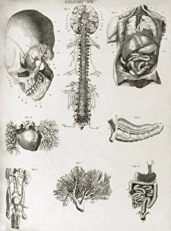
Background imageUrinary System Collection: Human anatomy, 1823 C017 / 8057

Human anatomy, 1823 C017 / 8057
Human anatomy. 19th-century illustrations showing the anatomy of a human skull (upper left), brain and spinal column (upper centre), chest and abdominal organs (lungs, heart, liver, stomach)

Background imageUrinary System Collection: Urinary system, artwork C015 / 6455

Urinary system, artwork C015 / 6455
Urinary system. Computer artwork showing the organs of the human urinary system. The kidneys (red, top left and right) filter waste products such as urea from the blood (red and blue) to form urine

Background imageUrinary System Collection: Urinary system, artwork C015 / 6454

Urinary system, artwork C015 / 6454
Urinary system. Computer artwork showing the organs of the human urinary system. The kidneys (red, top left and right) filter waste products such as urea from the blood to form urine

Background imageUrinary System Collection: Male anatomy, artwork C015 / 6448

Male anatomy, artwork C015 / 6448
Male anatomy. Computer artwork showing the major organs of the human torso. From top to bottom: oesophagus (white, top centre), trachea (windpipe, pink, top centre)

Background imageUrinary System Collection: Female anatomy, artwork C015 / 6447

Female anatomy, artwork C015 / 6447
Female anatomy. Computer artwork showing the major organs of the femaletorso. From top to bottom: oesophagus (white, top centre), trachea (windpipe, pink, top centre)

Background imageUrinary System Collection: Kidney stones, X-ray C017 / 7856

Kidney stones, X-ray C017 / 7856
Kidney stones. X-ray of the kidney of a patient with kidney stones (dark areas) in the renal pelvis (upper centre) of the kidney

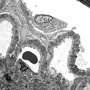
Background imageUrinary System Collection: Kidney glomerulus, TEM C016 / 5831

Kidney glomerulus, TEM C016 / 5831
Kidney glomerulus. Transmission electron micrograph (TEM) of a section through a renal glomerulus. At centre left is a capillary with a red blood cell (black) in its lumen

Background imageUrinary System Collection: Kidneys and ureters, artwork C013 / 4672

Kidneys and ureters, artwork C013 / 4672
Kidneys and ureters. Computer artwork showing an oblique anterior view of the kidneys, the main organs of the urinary system, relative to the skeleton and abdominal organs (white)

Background imageUrinary System Collection: Kidneys and ureters, artwork C013 / 4671

Kidneys and ureters, artwork C013 / 4671
Kidneys and ureters. Computer artwork showing an anterior view of the kidneys, the main organs of the urinary system, relative to the skeleton and abdominal organs (white)

Background imageUrinary System Collection: Urinary system, artwork C013 / 4673

Urinary system, artwork C013 / 4673
Urinary system. Computer artwork showing an anterior view of the kidneys, the main organs of the urinary system, relative to the skeleton and abdominal organs (white)

Background imageUrinary System Collection: Urinary system, artwork C013 / 4670

Urinary system, artwork C013 / 4670
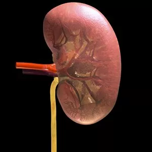
Urinary system. Computer artwork showing the kidneys (top), the main organs of the urinary system. The kidneys are located in the upper abdomen
Background imageUrinary System Collection: Urinary system, artwork C013 / 4669

Urinary system, artwork C013 / 4669
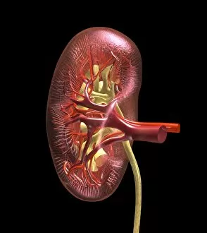
Urinary system. Computer artwork showing the kidneys (top), the main organs of the urinary system. The kidneys are located in the upper abdomen
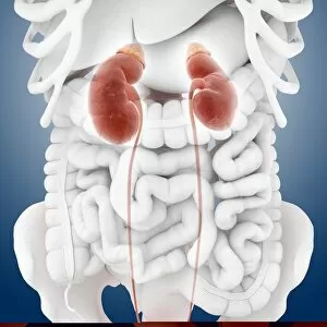
Background imageUrinary System Collection: Kidneys and ureters, artwork C013 / 4668

Kidneys and ureters, artwork C013 / 4668
Kidneys and ureters. Computer artwork showing the kidneys, the main organs of the urinary system. The kidneys are located in the upper abdomen

Background imageUrinary System Collection: Kidney tubule, TEM

Kidney tubule, TEM
Kidney tubule. Coloured transmission electron micrograph (TEM) of a section through a proximal convoluted tubule in the kidney

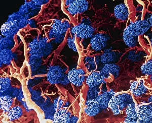
Background imageUrinary System Collection: Coloured SEM of glomeruli in the human kidney

Coloured SEM of glomeruli in the human kidney

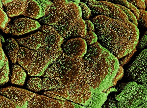
Background imageUrinary System Collection: Coloured SEM of healthy bladder epithelium

Coloured SEM of healthy bladder epithelium
Bladder epithelium. Coloured scanning electron micrograph (SEM) of healthy urinary bladder epithelium or mucosa. This lining is formed from epithelial cells (brown)


All Professionally Made to Order for Quick Shipping

The urinary system is a complex network of organs that play a vital role in maintaining the body's overall health and balance. From the intricate kidney tubules to the colorful MRI scan showcasing the kidneys and liver, this caption explores various aspects of this fascinating system. Starting with a closer look at kidney tubules in section, we delve into their microscopic structure and function. Moving on to an eye-catching color MRI scan of the abdomen, we witness the beauty of these essential organs alongside the liver. Shifting our focus to female anatomy, we encounter artwork depicting both the urinary bladder and a section through it. These illustrations provide insight into its unique composition and how it contributes to waste elimination. Zooming in even further, we explore kidney glomeruli through scanning electron microscopy (SEM). The detailed images reveal their intricate architecture responsible for filtering blood and producing urine. Additionally, SEM captures stunning visuals of kidney blood vessels intertwined within this remarkable filtration process. Further emphasizing their significance, light micrographs showcase multiple kidney glomeruli working harmoniously together. This glimpse highlights their collective effort towards maintaining fluid balance within our bodies. Concluding with captivating artwork portraying kidneys' overall anatomy, we gain a comprehensive understanding of these crucial organs' location and structure. Through this diverse collection of visuals ranging from scientific imaging techniques to artistic representations, one can truly appreciate the complexity and importance of our urinary system.