
Vascular Tissue Collection
Vascular tissue, the intricate network that ensures plants' survival, is a fascinating subject to explore


All Professionally Made to Order for Quick Shipping
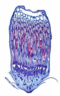
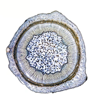
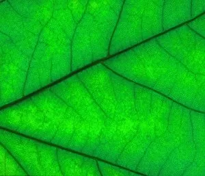
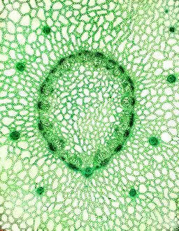
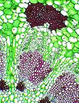
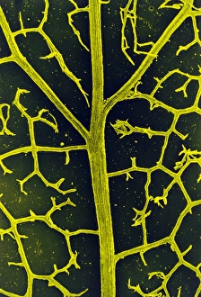
Vascular tissue, the intricate network that ensures plants' survival, is a fascinating subject to explore. Through light and scanning electron micrographs, we can witness its diverse forms and functions. In the lime tree stem's light micrograph, we observe xylem vessels transporting water and minerals from roots to leaves. These slender tubes act as nature's plumbing system, sustaining the entire plant. Similarly, in the water lily leaf's light micrograph, vascular tissue orchestrates nutrient distribution. It enables this aquatic beauty to thrive by efficiently absorbing resources from both water and soil. The dog rose stem showcases another aspect diversity. Its light micrograph reveals bundles of phloem cells responsible for transporting sugars produced during photosynthesis throughout the plant. Microscopic views of various plant tissues provide an insight into their complex structures. We marvel at how these tiny components work together harmoniously to support life processes within plants. Scanning electron microscopy exposes even more intricate details. The SEM image of xylem vessels unveils their elegant pattern resembling delicate lacework – a testament to nature's artistry. Another SEM image captures tobacco leaf B745/0312’s vascular tissue with remarkable clarity. This close-up view allows us to appreciate its intricacies on a cellular level while acknowledging its vital role in sustaining tobacco plants' growth. Wood also exhibits mesmerizing patterns when observed under SEM. The intricate arrangement of xylem cells provides strength and stability necessary for trees to withstand environmental challenges over time. A smilax root's light micrograph showcases yet another facet complexity – it highlights specialized cells adapted for efficient absorption of nutrients from surrounding soil particles. Lastly, multiple lime tree stem images emphasize its significance in understanding vascular tissue structure variations across different parts of a single plant organism - each serving specific purposes crucial for overall function and survival. Through dandelion flower's captivating light micrograph, we gain further appreciation for how even delicate structures possess vascular tissue, ensuring the plant's reproductive success.