Fraud Blocker Skip to main content

Vesicle Collection

"Exploring the Intricate World of Vesicles: Unveiling Cellular Secrets" Synapse nerve junction

Background imageVesicle Collection: Synapse nerve junction, TEM

Synapse nerve junction, TEM
Synapse. Coloured transmission electron micrograph (TEM) of a synapse, a junction between two nerve cells, in the brain. At a synapse an electrical signal is transmitted from one cell to the next in

Background imageVesicle Collection: Rough endoplasmic reticulum, TEM

Rough endoplasmic reticulum, TEM
Rough endoplasmic reticulum, coloured transmission electron micrograph (TEM). This section shows the rough endoplasmic reticulum (ER, folds, centre), a membranous structure that occurs in cells

Background imageVesicle Collection: Skin disorders, artwork

Skin disorders, artwork
Skin disorders. Computer artwork showing the structure of the skin, and the layers affected by various skin disorders. Folliculitis is the inflammation of hair follicles; Bullae are blisters;

Background imageVesicle Collection: Cell types, artwork

Cell types, artwork
Cell types. Cutaway artwork with a scale bar (upper right, in micrometres) showing the relative sizes of eukaryotic cells (those containing a nucleus) and prokaryotic cells (those lacking a nucleus)

Background imageVesicle Collection: Fibroblast cell, artwork

Fibroblast cell, artwork
Fibroblast cell. Computer artwork of a fibroblast excreting collagen fibres (tropocollagen). Fibroblasts are cells that produce connective tissue such as collagen (tropocollagen)

Background imageVesicle Collection: Golgi apparatus, SEM

Golgi apparatus, SEM
Golgi apparatus, coloured scanning electron micrograph (SEM). Section through a liver cell showing its Golgi apparatus (grey), a membrane-bound organelle that modifies and packages proteins

Background imageVesicle Collection: Nerve synapse, TEM

Nerve synapse, TEM
Nerve synapse. Coloured transmission electron micrograph (TEM) of the neuron (nerve) terminal at a synapse in the diaphragm

Background imageVesicle Collection: Bladderwrack, Fucus vesiculosus. Handcoloured woodblock engravings from James Main's Popular

Bladderwrack, Fucus vesiculosus. Handcoloured woodblock engravings from James Main's Popular Botany, Orr and Smith
FLO4671643 Bladderwrack, Fucus vesiculosus. Handcoloured woodblock engravings from James Main's Popular Botany, Orr and Smith, London, 1835

Background imageVesicle Collection: Development of the Tadpole (engraving)

Development of the Tadpole (engraving)
7188969 Development of the Tadpole (engraving) by European School, (19th century); Private Collection; (add.info.: Development of the Tadpole)

Background imageVesicle Collection: Bladderwrack, Fucus vesiculosus

Bladderwrack, Fucus vesiculosus. Handcoloured woodblock engravings from James Mains Popular Botany, Orr and Smith, London, 1835. James Main (1775-1846) was a Scottish gardener, botanist and writer

Background imageVesicle Collection: Eruptive fevers (colour litho)

Eruptive fevers (colour litho)
6005615 Eruptive fevers (colour litho) by English School, (19th century); Private Collection; (add.info.: Eruptive fevers)

Background imageVesicle Collection: Incubation of a chicken, Page from The Pictorial Museum of Animated Nature (engraving)

Incubation of a chicken, Page from The Pictorial Museum of Animated Nature (engraving)
979992 Incubation of a chicken, Page from The Pictorial Museum of Animated Nature (engraving) by English School, (19th century); Private Collection; (add.info.: chick growing in an egg)

Background imageVesicle Collection: Eruptive Fevers - Plate 6

Eruptive Fevers - Plate 6 - Measles, Scarlatina or Scarlet Fever, Vesicle of Vaccination (with slight surrounding rose rash), Typhus at an early stage

Background imageVesicle Collection: Shingles nerve damage

Shingles nerve damage

Background imageVesicle Collection: Operation a calculation of the gallbladder. Xilografia. Hort

Operation a calculation of the gallbladder. Xilografia. Hort
Medical history. Doctor operating a calculation of the gallbladder. Xilografia. Hortus Sanitatis by Johannes de Cuba (1430-1503). 15th C

Background imageVesicle Collection: Illustration of the structure of a plant cell, including nucleus, nucleolus, ribosome, endoplasmatic

Illustration of the structure of a plant cell, including nucleus, nucleolus, ribosome, endoplasmatic

Background imageVesicle Collection: Conceptual image of synaptic vesicles

Conceptual image of synaptic vesicles

Background imageVesicle Collection: Conceptual image of synapse receptors

Conceptual image of synapse receptors

Background imageVesicle Collection: Conceptual image of cytoskeleton

Conceptual image of cytoskeleton. The cytoskeleton is a cellular scaffolding or skeleton contained within a cells cytoplasm, and is present in all cells

Background imageVesicle Collection: Posterior view of urinary bladder showing seminal vesicles

Posterior view of urinary bladder showing seminal vesicles

Background imageVesicle Collection: Conceptual image of synaptic transmission

Conceptual image of synaptic transmission

Background imageVesicle Collection: The interior of an eukaryotic cell

The interior of an eukaryotic cell. This is the most common generic type of cell and its present in all mammals. In the center is the nucleus (the perforated sphere)

Background imageVesicle Collection: Clathrin Mediated Endocytosis

Clathrin Mediated Endocytosis

Background imageVesicle Collection: Scanning electron micrograph of an apoptotic HeLa cell

Scanning electron micrograph of an apoptotic HeLa cell. Zeiss Merlin HR-SEM

Background imageVesicle Collection: Bladder lining, TEM C014 / 1473

Bladder lining, TEM C014 / 1473
Bladder lining. Transmission electron micrograph (TEM) of a section through the epithelial cells that line of a human bladder, showing large flat vesicles (large, blue)

Background imageVesicle Collection: Nucleus and endoplasmic reticulum F006 / 9196

Nucleus and endoplasmic reticulum F006 / 9196
Computer artwork showing part of a human or eukaryotic cell. In the middle the nucleus which has a membrane with nuclear pores. Inside the nucleus is the DNA

Background imageVesicle Collection: Golgi apparatus, artwork F006 / 9211

Golgi apparatus, artwork F006 / 9211
Computer artwork of the Golgi apparatus of the human cell. This organelle functions as a central delivery system for the cell

Background imageVesicle Collection: Nucleus and endoplasmic reticulum F006 / 9201

Nucleus and endoplasmic reticulum F006 / 9201
Computer artwork showing part of a human or eukaryotic cell. In the middle the nucleus which has a membrane with nuclear pores. Inside the nucleus is the DNA

Background imageVesicle Collection: Golgi apparatus, artwork F006 / 9199

Golgi apparatus, artwork F006 / 9199
Computer artwork of the Golgi apparatus of the human cell. This organelle functions as a central delivery system for the cell

Background imageVesicle Collection: Golgi apparatus, artwork F006 / 9189

Golgi apparatus, artwork F006 / 9189
Computer artwork of the Golgi apparatus of the human cell. This organelle functions as a central delivery system for the cell

Background imageVesicle Collection: Legionella infected protozoan, TEM C016 / 9412

Legionella infected protozoan, TEM C016 / 9412
Legionella infected protozoan. Transmission electron micrograph (TEM) of a section through a single-celled Tetrahymena pyriformis protozoan, infected with Legionella pneumophila bacteria (yellow)

Background imageVesicle Collection: Stem cell dying, SEM

Stem cell dying, SEM
Stem cell dying. Coloured scanning electron micrograph (SEM) of a stem cell undergoing apoptosis, or programmed cell death. Apoptosis occurs when a cell becomes old or damaged

Background imageVesicle Collection: Golgi membranes, TEM

Golgi membranes, TEM
Golgi membranes. Transmission electron micrograph (TEM) of a section through a cell, showing the membranes (dark lines) of the Golgi apparatus

Background imageVesicle Collection: Enterocyte, TEM

Enterocyte, TEM
Enterocyte. Transmission electron micrograph (TEM) of a section through the cytoplasm and part of the nucleus of an enterocyte cell located in a crypt of Lieberkuhn of the small intestine

Background imageVesicle Collection: Clathrin lattice, molecular model C018 / 0453

Clathrin lattice, molecular model C018 / 0453
Clathrin lattice, molecular model. This polyhedral protein lattice coats eukaryotic cell membranes (vesicles) and is involved in protein secretion and membrane trafficking

Background imageVesicle Collection: Clathrin lattice, molecular model C018 / 0452

Clathrin lattice, molecular model C018 / 0452
Clathrin lattice, molecular model. This polyhedral protein lattice coats eukaryotic cell membranes (vesicles) and is involved in protein secretion and membrane trafficking

Background imageVesicle Collection: Clathrin lattice, molecular model C018 / 0454

Clathrin lattice, molecular model C018 / 0454
Clathrin lattice, molecular model. This polyhedral protein lattice coats eukaryotic cell membranes (vesicles) and is involved in protein secretion and membrane trafficking

Background imageVesicle Collection: Animal cell organelles, artwork

Animal cell organelles, artwork
Animal cell organelles. Artwork showing the organelles in a eukaryotic cell. This is an animal cell. Structures include the nucleus (centre) which has a membrane with nuclear pores (purple)

Background imageVesicle Collection: Nucleus and endoplasmic reticulum C015 / 6797

Nucleus and endoplasmic reticulum C015 / 6797
Computer artwork showing part of a human or eukaryotic cell. In the middle the nucleus which has a membrane with nuclear pores. Inside the nucleus is the DNA

Background imageVesicle Collection: Animal cell organelles, artwork C015 / 6793

Animal cell organelles, artwork C015 / 6793
Animal cell organelles. Computer artwork showing the organelles in a eukaryotic cell. This is an animal cell. Structures include the nucleus (centre) which has a membrane with nuclear pores

Background imageVesicle Collection: Animal cell organelles, artwork C015 / 6792

Animal cell organelles, artwork C015 / 6792
Animal cell organelles. Computer artwork showing the organelles in a eukaryotic cell. This is an animal cell. Structures include the nucleus (centre) which has a membrane with nuclear pores

Background imageVesicle Collection: Nucleus and endoplasmic reticulum C015 / 6783

Nucleus and endoplasmic reticulum C015 / 6783
Computer artwork showing part of a human or eukaryotic cell. In the middle the nucleus which has a membrane with nuclear pores. Inside the nucleus is the DNA

Background imageVesicle Collection: Nucleus and endoplasmic reticulum C015 / 6782

Nucleus and endoplasmic reticulum C015 / 6782
Computer artwork showing part of a human or eukaryotic cell. In the middle the nucleus which has a membrane with nuclear pores. Inside the nucleus is the DNA

Background imageVesicle Collection: Nucleus and endoplasmic reticulum C015 / 6781

Nucleus and endoplasmic reticulum C015 / 6781
Computer artwork showing part of a human or eukaryotic cell. In the middle the nucleus which has a membrane with nuclear pores. Inside the nucleus is the DNA

Background imageVesicle Collection: Animal cell organelles, artwork C015 / 6775

Animal cell organelles, artwork C015 / 6775
Animal cell organelles. Computer artwork showing the organelles in a eukaryotic cell. This is an animal cell. Structures include the nucleus (centre) which has a membrane with nuclear pores

Background imageVesicle Collection: Animal cell organelles, artwork C015 / 6778

Animal cell organelles, artwork C015 / 6778
Animal cell organelles. Computer artwork showing the organelles in a eukaryotic cell. This is an animal cell. Structures include the nucleus (centre) which has a membrane with nuclear pores

Background imageVesicle Collection: Animal cell organelles, artwork C015 / 6777

Animal cell organelles, artwork C015 / 6777
Animal cell organelles. Computer artwork showing the organelles in a eukaryotic cell. This is an animal cell. Structures include the nucleus (centre) which has a membrane with nuclear pores

Background imageVesicle Collection: Nucleus and endoplasmic reticulum C015 / 6772

Nucleus and endoplasmic reticulum C015 / 6772
Computer artwork showing part of a human or eukaryotic cell. In the middle the nucleus which has a membrane with nuclear pores. Inside the nucleus is the DNA


All Professionally Made to Order for Quick Shipping

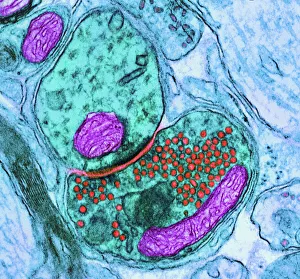
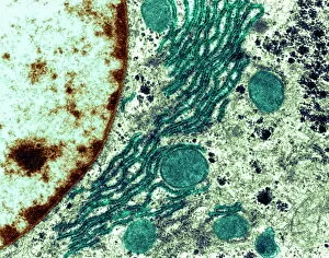
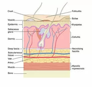
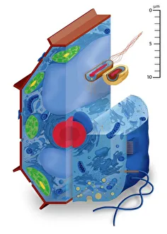
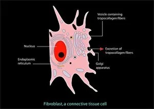
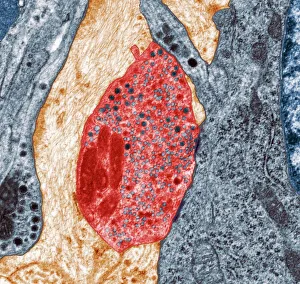
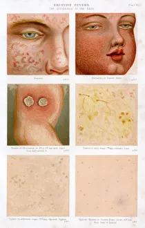
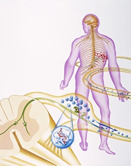
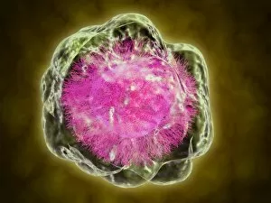
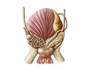
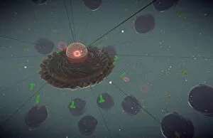
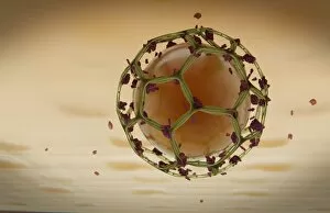
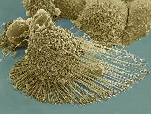
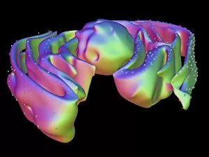
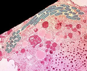
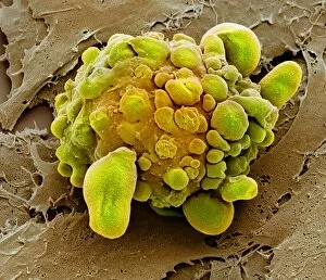
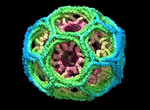
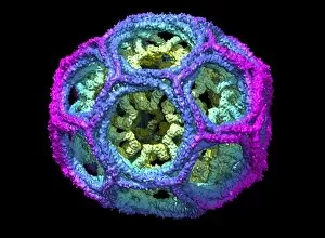
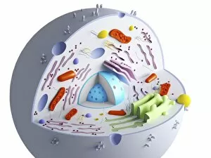
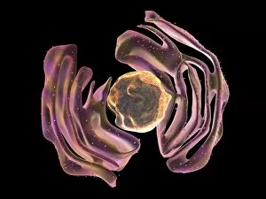
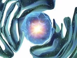
"Exploring the Intricate World of Vesicles: Unveiling Cellular Secrets" Synapse nerve junction, TEM: Witnessing the intricate communication between neurons at a synapse nerve junction through the lens of a transmission electron microscope (TEM). Rough endoplasmic reticulum, TEM: Peering into the rough endoplasmic reticulum within cells, revealing its role in protein synthesis and transport as captured by a transmission electron microscope (TEM). Skin disorders, artwork: Artistic representation showcasing various skin disorders that involve vesicle formation, highlighting their impact on human health and well-being. Cell types, artwork: A captivating artwork depicting different cell types where vesicles play crucial roles in cellular processes such as secretion and intracellular transport. Golgi apparatus, SEM: Exploring the intricacies of the Golgi apparatus using scanning electron microscopy (SEM), unraveling its vital function in modifying and packaging proteins within vesicles. Nerve synapse, TEM: Delving deep into the mesmerizing world of nerve synapses through high-resolution images obtained from a transmission electron microscope (TEM), shedding light on how vesicles facilitate neurotransmitter release. Fibroblast cell, artwork: An artistic portrayal capturing fibroblast cells involved in wound healing and collagen production while emphasizing their involvement with vesicular transport mechanisms. Development of Tadpole (engraving): A historical engraving illustrating tadpole development stages where they are instrumental in metamorphosis progression from aquatic to terrestrial lifeforms. Bladderwrack - Fucus vesiculosus: Discovering bladderwrack seaweed species like Fucus vesiculosus known for its distinctive air-filled reproductive structures called "vesicles, " which aid buoyancy and reproduction. Eruptive fevers - Colour litho & Plate 6 engraving .