Fraud Blocker Skip to main content

Zona Pellucida Collection

"Unlocking the Mystery of Fertilization

Background imageZona Pellucida Collection: Coloured SEM of egg & sperm during fertilisation

Coloured SEM of egg & sperm during fertilisation
Fertilisation. Coloured Scanning Electron Micro- graph (SEM) of a human egg (ovum) being penetrated by sperm during fertilisation. Sperm are coloured yellow, each with a rounded head and a long tail

Background imageZona Pellucida Collection: Ovarian follicle, SEM

Ovarian follicle, SEM
Ovarian follicle. Coloured scanning electron micrograph (SEM) of a fracture through a secondary follicle in the ovary. The oocyte (developing egg) is orange and its central nucleus is darker orange

Background imageZona Pellucida Collection: Ovarian follicle, light micrograph C016 / 0519

Ovarian follicle, light micrograph C016 / 0519
Ovarian follicle. Light micrograph of a section through a secondary ovarian follicle showing signs of maturation into the antral follicle class

Background imageZona Pellucida Collection: Sperm traveling towards egg with cellia

Sperm traveling towards egg with cellia

Background imageZona Pellucida Collection: Microscopic view of sperm traveling towards embryo

Microscopic view of sperm traveling towards embryo

Background imageZona Pellucida Collection: Microscopic view of ovules

Microscopic view of ovules

Background imageZona Pellucida Collection: Medical illustration of sperm and ovum

Medical illustration of sperm and ovum

Background imageZona Pellucida Collection: Conceptual image of common fertilization

Conceptual image of common fertilization

Background imageZona Pellucida Collection: Male reproductive sperm entering the egg

Male reproductive sperm entering the egg

Background imageZona Pellucida Collection: Microscopic view of sperm swimming towards egg

Microscopic view of sperm swimming towards egg

Background imageZona Pellucida Collection: Oocyte, light micrograph

Oocyte, light micrograph
Oocyte. Light micrograph of a section through an oocyte within a early antral follicle in an ovary. Oocytes are immature ova, or egg cells

Background imageZona Pellucida Collection: LM of four-cell embryo

LM of four-cell embryo
Four-cell embryo. Light micrograph of the blasto- meres of a four-cell human embryo, two days after fertilisation. The blastomeres are the rounded cells

Background imageZona Pellucida Collection: Coloured LM of a human embryo at two-cell stage

Coloured LM of a human embryo at two-cell stage

Background imageZona Pellucida Collection: Coloured LM of a fertilised human egg 24 hours old

Coloured LM of a fertilised human egg 24 hours old

Background imageZona Pellucida Collection: SEM of ovulation; release of egg

SEM of ovulation; release of egg

Background imageZona Pellucida Collection: LM of blastomeres of a two-cell embryo

LM of blastomeres of a two-cell embryo
Two-cell embryo. Light micrograph of the blasto- meres of a 2-cell human embryo, two days after fertilisation. The blastomeres are the rounded cells formed by the first division of the fertilized

Background imageZona Pellucida Collection: Coloured SEM showing the ovulation process

Coloured SEM showing the ovulation process
Ovulation. Coloured scanning electron micrograph of the ovulation process. The egg (red at centre) has ruptured the external surface of the ovary (dark green), known as the germinal epithelium

Background imageZona Pellucida Collection: Artwork of a human egg cell

Artwork of a human egg cell in the primordial follicle phase which occurs after seven months of fetal development. The egg cell is surrounded by a thick layer of follicular cells known as zona

Background imageZona Pellucida Collection: Sperm fertilising an egg, artwork

Sperm fertilising an egg, artwork
Sperm fertilising an egg. Cutaway artwork of a human sperm cell (spermatozoon) penetrating an egg cells (ovum) thick outer layer (zona pellucida). The egg cells membrane is at top right

Background imageZona Pellucida Collection: Fertilisation, computer artwork

Fertilisation, computer artwork
Fertilisation. Computer artwork of a sperm cell (purple) penetrating a human egg (ovum). Each sperm (spermatozoan) has a rounded head (acrosome) and a long tail with which it swims

Background imageZona Pellucida Collection: Fertilization

Fertilization. Artwork of sperm cells (white and black) surrounding a human egg cell (round). The egg cell is protected by an outer layer, the zona pellucida (black)

Background imageZona Pellucida Collection: Ovarian follicle, SEM

Ovarian follicle, SEM
Ovarian follicle. Coloured scanning electron micrograph (SEM) of a fracture through a secondary follicle in the ovary. The oocyte (developing egg), with its central nucleus, is purple

Background imageZona Pellucida Collection: Ovarian follicle maturation, artwork

Ovarian follicle maturation, artwork
Ovarian follicle maturation. Computer artwork showing the beginning of the follicular phase of ovarian follicle maturation. At bottom left is the primary follicle stage

Background imageZona Pellucida Collection: Fertilisation, artwork

Fertilisation, artwork
Fertilisation. Computer artwork of a sperm cell (blue) penetrating a human egg (ovum). Each sperm (spermatozoan) has a rounded head (acrosome) and a long tail with which it swims


All Professionally Made to Order for Quick Shipping

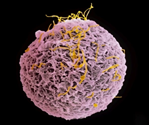
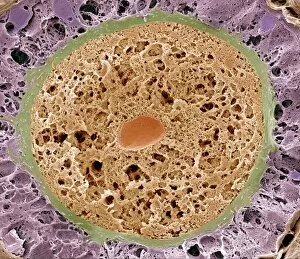
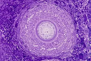
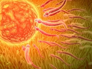
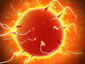
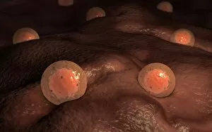
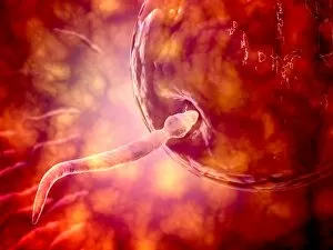
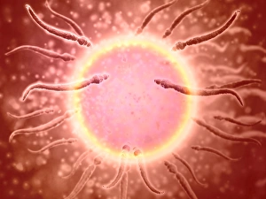
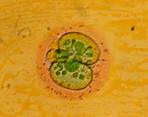
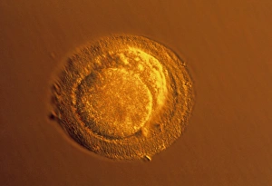
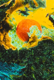
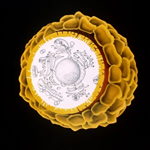
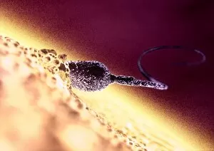
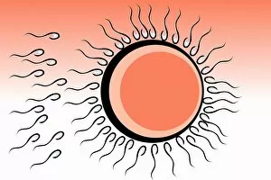
"Unlocking the Mystery of Fertilization: Exploring the Zona Pellucida" Witness the mesmerizing dance of life as a colored SEM captures the moment of fertilization between an egg and sperm. Delve into the intricate world within ovarian follicles through a stunning SEM image, revealing their role in reproduction. A captivating light micrograph C016/0519 showcases the delicate beauty of an ovarian follicle, hinting at its crucial function in fertility. Marvel at nature's design as sperm with cellia propel themselves towards an awaiting egg, captured in a microscopic view that leaves you in awe. Embark on a microscopic journey witnessing sperm swimming tirelessly towards their destination - an embryo ready to be formed. Witness the magical release of eggs from female ovaries, depicted through a vivid illustration that unveils this pivotal event in reproduction. Dive deeper into nature's wonders with another microscopic view showcasing determined sperm making their way towards an eagerly anticipated egg. Explore ovules up close through multiple microscope views, unraveling their significance in creating new life and continuing generations. Discover medical illustrations depicting both sperm and ovum, offering insights into their unique structures and how they unite to create life itself. Immerse yourself once again in a breathtaking microscopic view capturing the relentless journey of sperm swimming passionately towards their destined union with an egg. In this conceptual image portraying common fertilization, we are reminded of nature's incredible ability to perpetuate life through this miraculous process called "zona pellucida.