
Hepatology Collection
Hepatology: Exploring the Complexities of Liver Health and Disease The liver, a vital organ responsible for numerous essential functions in our body


All Professionally Made to Order for Quick Shipping
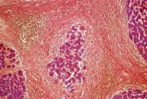
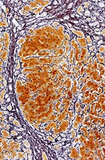
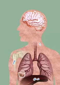
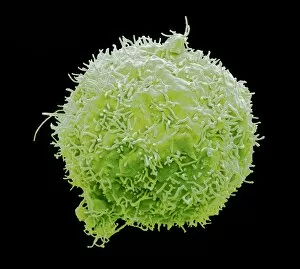
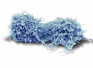
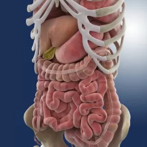
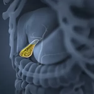
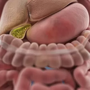
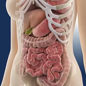
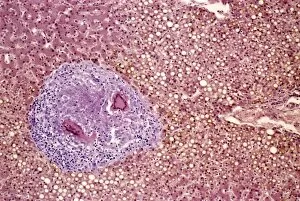
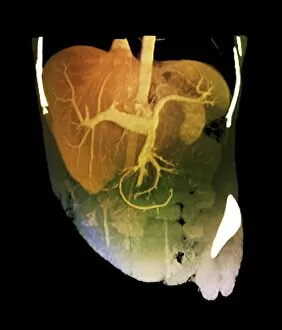
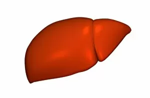
Hepatology: Exploring the Complexities of Liver Health and Disease The liver, a vital organ responsible for numerous essential functions in our body, can face various challenges that affect its health. One such condition is liver tissue cirrhosis, which can be observed through a light micrograph. This image showcases the structural changes within the liver caused by chronic damage and scarring. Intriguingly, another aspect involves studying dividing liver cancer cells under scanning electron microscopy (SEM). These detailed images provide valuable insights into the cellular processes associated with this devastating disease. The SEM captures the intricate division of liver cancer cells, highlighting their abnormal growth patterns. Metastatic cancer is yet another area explored within hepatology. Through captivating artwork representations, we gain an understanding of how cancerous cells spread from their primary site to infiltrate other parts of the body. These illustrations depict both the complexity and urgency surrounding metastatic liver cancer research. Delving deeper into liver anatomy, artistic renditions offer a comprehensive view of this remarkable organ's structure and function. Artwork like C016 / 7001 provides an educational glimpse into different lobes and vessels that make up our complex hepatic system. Returning to SEM imagery, we witness further exploration into liver cancer cells' characteristics. Multiple high-resolution scans reveal distinct features specific to these malignant cells – irregular shapes, abnormal sizes – all contributing to better identification and understanding for researchers working tirelessly towards effective treatments. As hepatologists continue their pursuit in unraveling mysteries surrounding diseases like cirrhosis or developing innovative therapies for combating metastatic cancers affecting the liver; these captivating visuals serve as reminders of both challenges faced and progress achieved in this critical field.