
Uterus Collection
"The Wondrous Uterus: A Journey of Life and Creation" Delve into the intricate world of the uterus through pen and ink studies by Leonardo da Vinci, circa 1510


All Professionally Made to Order for Quick Shipping
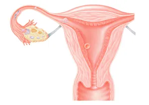
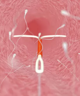
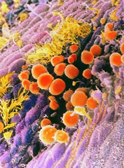
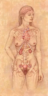
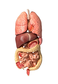
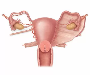
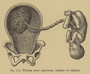
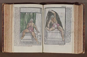
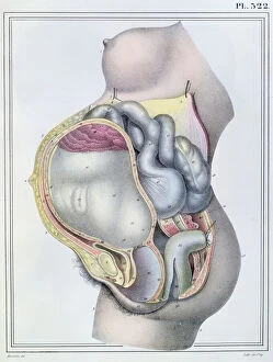
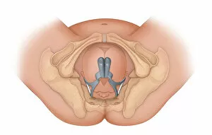
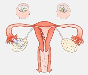
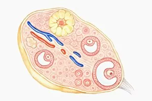
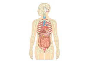
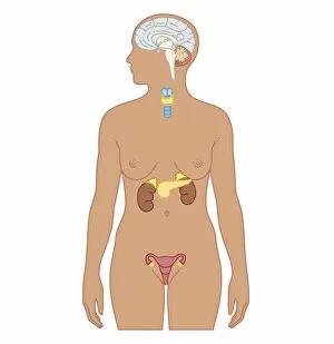
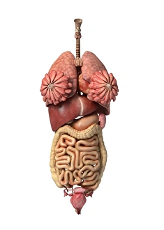
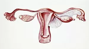
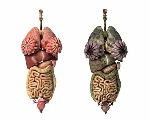
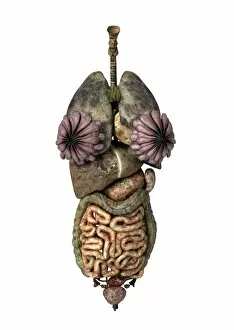
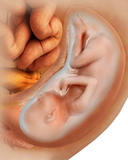
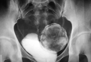
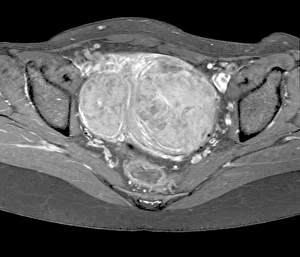
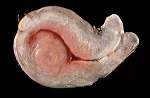
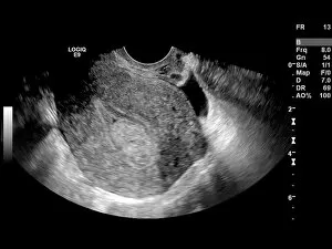
"The Wondrous Uterus: A Journey of Life and Creation" Delve into the intricate world of the uterus through pen and ink studies by Leonardo da Vinci, circa 1510. These captivating illustrations offer a glimpse into the early understanding of human fetal development. Witness the remarkable transformation within the uterus as you explore scanning electron microscope images showcasing the delicate lining during menstruation. Marvel at its resilience and ability to shed, preparing for new beginnings. Embark on a digital illustration depicting the awe-inspiring journey of a fertilized human egg. Follow its path towards implantation, where it finds solace in the nurturing embrace of this miraculous organ. Discover modern contraceptive methods like IUDs alongside microscopic views of sperm cells, highlighting their interaction with this vital reproductive structure. Witness how science empowers individuals to make choices about their own bodies. Step inside an amniotic sac housing a developing human fetus, encapsulating life's earliest stages within this protective sanctuary. Experience firsthand nature's masterpiece unfolding before your eyes. Travel back in time through a late 19th-century wood engraving that intricately depicts the female reproductive system. Admire its complexity and marvel at how far our knowledge has come since then. Observe uterine cells during childbirth – witness their strength as they contract and facilitate new life entering into this world. Celebrate women's incredible capacity for creation and nurture. Immerse yourself in vivid color scanning electron microscope images capturing every detail of the uterine lining or endometrium – an essential component ensuring fertility and supporting pregnancy. Uncover biomedical illustrations offering cross-sectional insights into the female reproductive system - admire its intricacies while gaining appreciation for its role in sustaining life itself. Explore front view anatomical renderings highlighting not only physical structures but also emphasizing connections with an intricate endocrine system.