Jigsaw Puzzle : Teenager hand X-ray


![]()
Jigsaw Puzzles from Science Photo Library
Teenager hand X-ray
Teenagers hand bones (13 year old). Coloured X- ray showing the bones in the hand of a thirteen year old teenager. Bones appear white. At this age not much cartilage is seen between each bone joint and ossification (bone formation) has occurred widely so that the spaces between the joints have narrowed. All wrist bones have formed and are visible at centre left. This hand is close to the adult bone pattern. At far left are the radius and ulna bones of the forearm
Science Photo Library features Science and Medical images including photos and illustrations
Media ID 6448643
© CNRI/SCIENCE PHOTO LIBRARY
Bones Child Finger Bone Growth Hand Ossification Phalanges Tee Nager
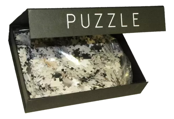
Jigsaw Puzzle (500 Pieces)
Discover the wonders of the human body with our captivating Teenager Hand X-ray Jigsaw Puzzle from Media Storehouse, in partnership with Science Photo Library. This intriguing puzzle showcases the vibrant colors of a thirteen-year-old teenager's hand bones as revealed in this X-ray image. Assemble this challenging puzzle to deepen your understanding of anatomy and inspire curiosity about the intricacies of the human body. Perfect for individuals of all ages and skill levels, this puzzle promises an engaging and educational experience.
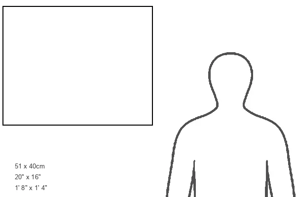
500 piece puzzles are custom made in Australia and hand-finished on 100% recycled 1.6mm thick laminated puzzle boards. There is a level of repetition in jigsaw shapes with each matching piece away from its pair. The completed puzzle measures 40x51cm and is delivered packaged in an attractive presentation box specially designed to fit most mail slots with a unique magnetic lid
Jigsaw Puzzles are an ideal gift for any occasion
Estimated Product Size is 50.7cm x 40.3cm (20" x 15.9")
These are individually made so all sizes are approximate
Artwork printed orientated as per the preview above, with landscape (horizontal) or portrait (vertical) orientation to match the source image.
EDITORS COMMENTS
This print showcases the intricate structure of a thirteen-year-old teenager's hand bones. The colored X-ray reveals a close-up view of the hand, with each bone appearing white against the dark background. At this stage of development, not much cartilage is visible between the joints, indicating significant ossification or bone formation. The image highlights how bone growth and development progress during adolescence. The spaces between the joints have narrowed as ossification has occurred widely throughout the hand. All wrist bones are clearly formed and can be seen at the center-left portion of the X-ray. Interestingly, this particular hand closely resembles an adult bone pattern, demonstrating that skeletal maturity is approaching. On the far left side of the image, we can observe both radius and ulna bones from the forearm. This photograph provides valuable insights into human anatomy, specifically focusing on skeleton growth in teenagers. It emphasizes how bones evolve over time and offers a glimpse into their complex structure at different stages of life. Overall, this visually striking image captures both scientific curiosity and aesthetic appeal while shedding light on topics such as bone development, ossification processes, and finger phalanges in young individuals.
MADE IN AUSTRALIA
Safe Shipping with 30 Day Money Back Guarantee
FREE PERSONALISATION*
We are proud to offer a range of customisation features including Personalised Captions, Color Filters and Picture Zoom Tools
SECURE PAYMENTS
We happily accept a wide range of payment options so you can pay for the things you need in the way that is most convenient for you
* Options may vary by product and licensing agreement. Zoomed Pictures can be adjusted in the Cart.