Fraud Blocker Skip to main content

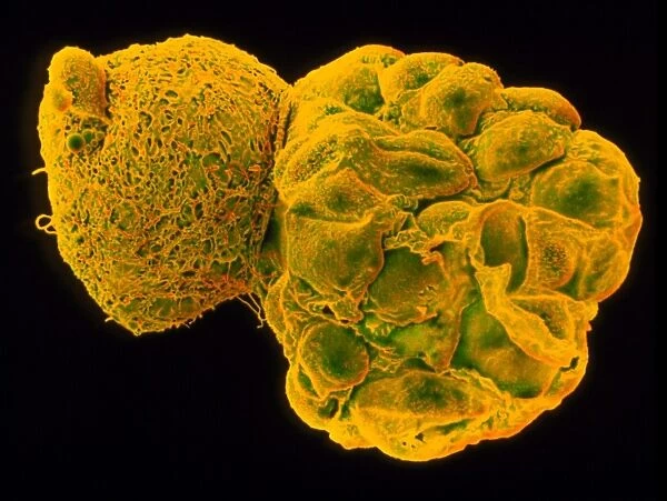
Coloured SEM of a hatching blastocyst 5 days old

Coloured SEM of a hatching blastocyst 5 days old
In Stock
We accept all major payment options so you can pay for the things you need in the way that is most convenient for you
Framed Photos
Buy from AU$119.99
Photo Prints
Buy from AU$12.99
Jigsaw Puzzles
Buy from AU$89.99
Poster Prints
Buy from AU$24.99
Canvas Prints
Buy from AU$124.99
Fine Art Prints
Buy from AU$92.53
Metal Prints
Buy from AU$43.99
Cards
Buy from AU$12.99
Home Decor
Buy from AU$24.99

Science Photo Library Photo Prints and Wall Art

Coloured SEM of a hatching blastocyst 5 days old

Hatching blastocyst embryo. Coloured scanning electron micrograph (SEM) of a human embryo at the blastocyst stage, five days after fertilisation. It is seen at right hatching from a hole in the zona pellucida (at left), a protein shell that originally surrounded the unfertilised egg. The blastocyst is a hollow ball of cells with a fluid centre. Each cell is called a blastomere. Most of these embryo cells will form the placenta and membranes around the embryo, and only a small group (the inner mass) form the embryo proper. At this stage the blastocyst has moved into the uterus (womb) and is preparing to implant on the womb wall. Magnification: x360 at 6x7cm size. Magnification; x1, 200 at 8x10 inch size. Science Photo Library features Science and Medical images including photos and illustrations. © DR YORGOS NIKAS/SCIENCE PHOTO LIBRARY

Media ID 6453841

Blastocyst Embryo Hatching

SECURE PAYMENTS
We accept all major payment options so you can pay for the things you need in the way that is most convenient for you

Made in Australia
Safe Shipping, Easy Returns

FREE PERSONALISATION
We are proud to offer a range of customisation including Cropping, Captions, and Filters. Zoom Cropping can be adjusted in the basket


EDITORS COMMENTS

This print showcases the intricate beauty of life at its earliest stages. Captured through a scanning electron microscope, it features a coloured SEM image of a hatching blastocyst embryo that is five days old. The blastocyst, seen on the right side of the image, is emerging from a small opening in the zona pellucida—a protective protein shell that once enveloped the unfertilised egg. The blastocyst itself resembles a hollow ball composed of numerous cells known as blastomeres. Remarkably, most of these cells will go on to form vital components such as the placenta and membranes surrounding the developing embryo. Only a select group called the inner mass will eventually give rise to what we recognize as an embryo. At this critical stage, approximately five days after fertilisation has taken place, the blastocyst has migrated into the uterus and is preparing for implantation onto its wall—an essential step in pregnancy initiation. With magnification levels reaching x360 at 6x7cm size or x1,200 at 8x10 inch size, this visually stunning photograph offers us an extraordinary glimpse into our own beginnings—the miraculous journey from conception to new life.


Framed Prints of Coloured SEM of a hatching blastocyst 5 days old

Bring the wonders of science into your home with Media Storehouse's Framed Prints. This captivating coloured Scanning Electron Micrograph (SEM) image, sourced from Science Photo Library, showcases a human embryo at the blastocyst stage, just five days after fertilisation. Witness the intricacy of life's earliest moments with this stunning, high-definition print. A perfect addition to any scientific or educational space, this framed print is sure to ignite curiosity and inspire wonder.


Photo Prints of Coloured SEM of a hatching blastocyst 5 days old

Discover the wonders of life's earliest stages with Media Storehouse's range of Photographic Prints. This captivating image showcases a coloured Scanning Electron Micrograph (SEM) of a human embryo at the blastocyst stage, just five days after fertilisation. Witness the intricate details of the hatching blastocyst as it emerges, bringing the marvels of science and human development to your home or office. A must-have for scientists, educators, or anyone with a curiosity for the miracles of life.


Poster Prints of Coloured SEM of a hatching blastocyst 5 days old

Discover the wonders of embryonic development with our Media Storehouse Poster Prints. Featuring an awe-inspiring coloured Scanning Electron Micrograph (SEM) image of a hatching blastocyst, five days old, by Science Photo Library. This captivating print offers a unique and intriguing glimpse into the early stages of human life. Bring the magic of science into your home or office with this educational and visually stunning poster print.


Jigsaw Puzzles of Coloured SEM of a hatching blastocyst 5 days old

Discover the wonders of embryonic development with Media Storehouse's Jigsaw Puzzles. This intriguing puzzle showcases a coloured Scanning Electron Micrograph (SEM) of a hatching blastocyst, five days old, captured by Science Photo Library. With stunning detail and vibrant colours, this puzzle offers a unique and educational experience. Challenge yourself and your family to piece together the complex structure of this early embryo, revealing the intricacies of human development. A perfect addition to any home or classroom, this puzzle is sure to inspire curiosity and learning.

diamond
Made With High Grade Materials
thumb_up
Professional Quality Finish
package_2
Carefully Packed To Aid Safe Arrival

You May Also Like


The Dom Cathedral and the River RhineThe Dom Cathedral and the River Rhine
Add to Basket
Locomotive cleaners, about 1916Locomotive cleaners, about 1916
Add to Basket
Portal vein surgery, X-ray C016 / 6543Portal vein surgery, X-ray C016 / 6543
Add to Basket
People in Highgate Woods, North LondonPeople in Highgate Woods, North London
Add to Basket
Al Gore at Stonehenge DP137787Al Gore at Stonehenge DP137787
Add to Basket
Congressional Club, between 1917 and 1920. Creator: Harris & Ewing. Congressional Club, between 1917 and 1920. Creator: Harris & EwingCongressional Club, between 1917 and 1920. Creator: Harris & Ewing. Congressional Club, between 1917 and 1920. Creator: Harris & Ewing
Add to Basket

Gallery Wall Inspiration

Nerve cell, SEM
False-coloured SEM (Scanning Electron Micrograph) of a Tardigrade (Tardigrada) amongst moss, from Peak District National Park, Derbyshire, UK. This tardigrade is approximately 0. 1 millimetres long. Digital composite
Santos-Dumont / Sem
Neutrophil engulfing MRSA, SEM C018 / 8596
X and Y chromosomes
Lavender pollen grain, SEM
Scanning Electron Micrograph (SEM): Tardigrade or ‘Water Bear Magnification x 1250 (A4 size: 29. 7 cm width)
Cat tongue surface, SEM
Hazel pollen grains
E. coli bacteria, SEM
Collagen, Scanning electron micrograph (SEM)
Scanning electron micrograph (SEM) of red blood cell
T lymphocytes and cancer cell, SEM C001 / 1679
Tardigrade or Water Bear
Crysotile asbestos
Uterus lining during menstruation, SEM
Iris of the eye, SEM
Stylus in LP record
Macrophage engulfing TB bacteria, SEM
Salmonella bacteria, SEM
SEM of penicillin fungus
Vascular bundle, SEM
Geranium anther, SEM
Dahlia flower pollen, SEM
Scanning Electron Micrograph (SEM): Praying Mantis - Magnification x 30 (if print A4 size: 29. 7 cm wide)
Tardigrade, SEM
Botanik Digitalis purpurea L. Fingerhut 160: 1


© Copyright Media Storehouse, All Rights Reserved 2003 - 2024
Facebook     Pinterest     100% Payment Secure     Reviews IO