Fraud Blocker Skip to main content

Home > Science Photo Library > Images Dated > 2008 > April > 16 Apr 2008

Images Dated 16th April 2008

Choose a picture from our Images Dated 16th April 2008 Collection for your Wall Art and Photo Gifts

40 items

We are proud to offer this selection in partnership with Science Photo Library

Background imageImages Dated 16th April 2008: Magnetic field

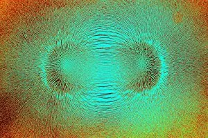
Magnetic field. Bar magnet with iron filings aligned around it. The magnetic field induces magnetism in each of the filings, which then line up in the field

Background imageImages Dated 16th April 2008: Heathland ants antenna, SEM

Heathland ants antenna, SEM
Ants antenna. Coloured scanning electron micrograph (SEM), of sensory hairs on the antenna of a heathland ant (Formica fusca). Magnification: x400 when printed at 10 centimetres wide

Background imageImages Dated 16th April 2008: Mole hair, SEM

Mole hair, SEM
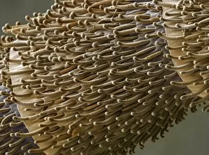
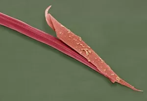
Mole hair. Coloured scanning electron micrograph (SEM) of a skin of a mole (family Talpidae), showing fine hairs. Moles are small mammals that live in underground burrows

Background imageImages Dated 16th April 2008: Daubentons bat hairs, SEM

Daubentons bat hairs, SEM
Bat hairs. Coloured scanning electron micrograph (SEM) of hairs from Daubentons bat (Myotis daubentonii). Each hair shaft has an outer cuticle made up of overlapping scales, or plates

Background imageImages Dated 16th April 2008: Olive leaf trichomes, SEM

Olive leaf trichomes, SEM
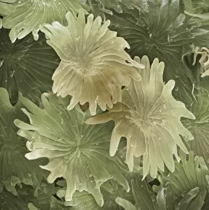
Olive leaf trichomes. Coloured scanning electron micrograph (SEM) of peltate trichomes, or modified hairs, on the surface of an olive leaf (Olea europaea)

Background imageImages Dated 16th April 2008: Light bulb filament, SEM

Light bulb filament, SEM
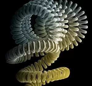
Tungsten light bulb filament, coloured scanning electron micrograph (SEM). The heat generated by an electrical current flowing through the filament makes it glow white hot

Background imageImages Dated 16th April 2008: Household dust, SEM

Household dust, SEM
Household dust, coloured scanning electron micrograph (SEM). Magnification: x130 when printed at 10 centimetres wide

Background imageImages Dated 16th April 2008: Marrow pollen grain, SEM

Marrow pollen grain, SEM
Marrow (Cucurbita sp.) pollen grain, scanning electron micrograph (SEM). The grain has a spiked outer wall (exine), which may assist in its dispersal by improving its adherence to pollinating

Background imageImages Dated 16th April 2008: Olive leaf trichomes, SEM

Olive leaf trichomes, SEM
Olive leaf trichomes. Coloured scanning electron micrograph (SEM) of peltate trichomes, or modified hairs, on the surface of an olive leaf (Olea europaea)

Background imageImages Dated 16th April 2008: Head louse, SEM

Head louse, SEM
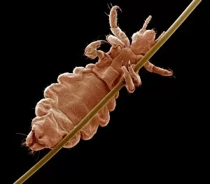
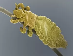
Head louse. Coloured scanning electron micrograph (SEM) of a head louse (Pediculus humanus capitis) clinging to a human hair. Head lice measure 2-3 millimetres in length

Background imageImages Dated 16th April 2008: Foraminiferan, SEM

Foraminiferan, SEM
Foraminiferan. Coloured scanning electron micrograph (SEM) of a foraminiferan fossil. Foraminiferans are marine single-celled protists that construct and inhabit shells (tests)

Background imageImages Dated 16th April 2008: Head louse, SEM

Head louse, SEM
Head louse. Coloured scanning electron micrograph (SEM) of a head louse (Pediculus humanus capitis) clinging to a human hair. Head lice measure 2-3 millimetres in length

Background imageImages Dated 16th April 2008: Head louse, SEM

Head louse, SEM
Head louse. Coloured scanning electron micrograph (SEM) of a head louse (Pediculus humanus capitis) clinging to a human hair. Head lice measure 2-3 millimetres in length

Background imageImages Dated 16th April 2008: Head louse, SEM

Head louse, SEM
Head louse. Coloured scanning electron micrograph (SEM) of a head louse (Pediculus humanus capitis) clinging to a human hair. Head lice measure 2-3 millimetres in length

Background imageImages Dated 16th April 2008: Nematode worms, SEM

Nematode worms, SEM
Nematode worm. Coloured scanning electron micrograph (SEM) of a juvenile Heterorhabditis bacteriophora nematode worm (centre) shedding its cuticle (outer coat)

Background imageImages Dated 16th April 2008: Head louse, SEM

Head louse, SEM
Head louse. Coloured scanning electron micrograph (SEM) of the head of a head louse (Pediculus humanus capitis). Its mouthparts are at centre. Head lice measure 2-3 millimetres in length

Background imageImages Dated 16th April 2008: Nematode worms, SEM

Nematode worms, SEM
Nematode worm. Scanning electron micrograph of a juvenile Heterorhabditis bacteriophora nematode worm (centre) shedding its cuticle (outer coat, centre right) as it develops into an adult

Background imageImages Dated 16th April 2008: Foraminiferan, SEM

Foraminiferan, SEM
Foraminiferan. Coloured scanning electron micrograph (SEM) of a foraminiferan fossil. Foraminiferans are marine single-celled protists that construct and inhabit shells (tests)

Background imageImages Dated 16th April 2008: Lacewing larva, SEM

Lacewing larva, SEM
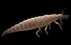
Lacewing larva (Chrysoperla carnea), coloured scanning electron micrograph (SEM). This insect preys on aphids, whitefly and other insects

Background imageImages Dated 16th April 2008: Mosquito heads, light micrograph

Mosquito heads, light micrograph
Mosquito heads. Light micrograph of the heads of a male (left) and female (right) mosquito (family Culicidae). The mosquitos head is dominated by its large compound eyes (solid white)

Background imageImages Dated 16th April 2008: Aphids, SEM

Aphids, SEM
Aphids. Coloured scanning electron micrograph (SEM) of aphids (family Aphididae), or greenflies, feeding on a plant stem. Only their abdomens can be seen here

Background imageImages Dated 16th April 2008: Mosquito heads, light micrograph

Mosquito heads, light micrograph
Mosquito heads. Light micrograph of the heads of a male (left) and female (right) mosquito (family Culicidae). The mosquitos head is dominated by its large compound eyes (black spheres)

Background imageImages Dated 16th April 2008: Nose mucosa, light micrograph

Nose mucosa, light micrograph
Nose mucosa. Light micrograph of a section through the nasal mucosa, the tissue that lines the airways of the nose. Air flows through hair-like cilia

Background imageImages Dated 16th April 2008: Ovarian follicle, light micrograph

Ovarian follicle, light micrograph
Ovarian follicle. Coloured light micrograph of a section through a secondary follicle in an ovary. At upper left is the large nucleus of the developing egg (oocyte, green)

Background imageImages Dated 16th April 2008: Ovarian follicle, light micrograph

Ovarian follicle, light micrograph
Ovarian follicle. Coloured light micrograph of a section through a secondary follicle in an ovary. At upper left is the large nucleus of the developing egg (oocyte, orange)

Background imageImages Dated 16th April 2008: Lymph node, light micrograph

Lymph node, light micrograph
Lymph node. Coloured light micrograph of a section through a lymph node. A lymph node filters pathogens from lymph fluid, a watery liquid that surrounds the tissues of the body

Background imageImages Dated 16th April 2008: Lymph node, light micrograph

Lymph node, light micrograph
Lymph node. Coloured light micrograph of a section through a lymph node. A lymph node filters pathogens from lymph fluid, a watery liquid that surrounds the tissues of the body

Background imageImages Dated 16th April 2008: Aristotle as a young man

Aristotle as a young man. The Ancient Greek philosopher and naturalist Aristotle (384-322 BC) influenced many later thinkers, working in areas such as logic, astronomy, zoology

Background imageImages Dated 16th April 2008: Claude Bernard, French physiologist

Claude Bernard, French physiologist
Claude Bernard (1813-1878), French physiologist and pioneer of experimental medicine and physiological chemistry. Bernards discoveries were wide-ranging

Background imageImages Dated 16th April 2008: Claude Louis Berthollet, French chemist

Claude Louis Berthollet, French chemist
Claude-Louis Berthollet (1748-1822), French chemist. Berthollet was an early supporter of Lavoisier, but correctly disagreed with him by proposing that some acids do not contain oxygen

Background imageImages Dated 16th April 2008: Archimedes, Greek mathematician

Archimedes, Greek mathematician
Archimedes of Syracuse (c. 287-212 BC), Greek mathematician, physicist and engineer. Archimedes is regarded as the greatest mathematician of ancient times

Background imageImages Dated 16th April 2008: Archimedes, Greek mathematician

Archimedes, Greek mathematician
Archimedes of Syracuse (c. 287-212 BC), Greek mathematician, physicist and engineer. Archimedes is regarded as the greatest mathematician of ancient times

Background imageImages Dated 16th April 2008: George Biddell Airy, British astronomer

George Biddell Airy, British astronomer
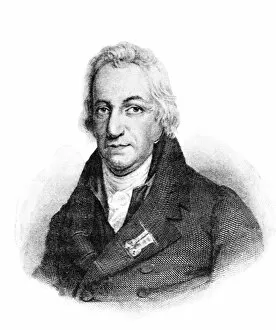
George Biddell Airy (1801-1892), British astronomer, 19th century engraving. During his career, Airy was elected president to the Royal Astronomical Society

Background imageImages Dated 16th April 2008: Tooth fossil, SEM

Tooth fossil, SEM
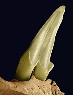
Tooth fossil. Coloured scanning electron micrograph (SEM) of a fossilised canine tooth of the extinct Puerto Rican shrew (Nesophontes edithae)

Background imageImages Dated 16th April 2008: White lace flower seed, SEM

White lace flower seed, SEM
White lace flower seed. Coloured scanning electron micrograph (SEM) of a seed from a white lace flower (Orlaya sp.). A seed contains an embryo (a fertilised ovule from which a new plant can grow)

Background imageImages Dated 16th April 2008: Euphorbia seeds, SEM

Euphorbia seeds, SEM
Euphorbia seeds. Coloured scanning electron micrograph (SEM) of seeds from a Euphorbia flower (Euphorbia sp.). A seed contains an embryo (a fertilised ovule from which a new plant can grow)

Background imageImages Dated 16th April 2008: St. Johns wort seeds, SEM

St. Johns wort seeds, SEM
St. Johns wort seeds. Coloured scanning electron micrograph (SEM) of seeds from a St. Johns wort flower (Hypericum sp.). A seed contains an embryo (a fertilised ovule from which a new plant can grow)

Background imageImages Dated 16th April 2008: White lace flower seed, SEM

White lace flower seed, SEM
White lace flower seed. Coloured scanning electron micrograph (SEM) of a seed from a white lace flower (Orlaya sp.). A seed contains an embryo (a fertilised ovule from which a new plant can grow)

Background imageImages Dated 16th April 2008: Fungal hyphae, SEM

Fungal hyphae, SEM
Fungal hyphae. Coloured scanning electron micrograph (SEM) of the hyphae of a fungus. Hyphae are thread-like structures that form the mycelium


All Professionally Made to Order for Quick Shipping


-

Why Choose Us?

We are a leading provider of Art Prints and Photo Gifts since 2003, working in partnership with a range of Sporting Clubs, Charities, Museums and Picture Libraries. A large share of profits from any of their images will go directly towards supporting that charity or club. Our archive of images is carefully curated to bring you a wide range of subjects, including landscapes, wildlife, architecture, and more. We ship from our partner labs in the UK, USA, EU (Netherlands) and Australia.
+

How do I place an order?

Ordering is quick & easy - Just follow 5 Simple Steps:
  1. Find your image: Use our search box or browse our online photo Collections to find the image you want.

  2. Choose your print format: Select your desired product and add it to your cart.

  3. Enter your details: If you're a returning customer, simply enter your email address and password, and we'll fill in your billing and shipping address details. All personal details are held securely and are fully GDPR compliant. As standard, we remove all Personally Identifiable Information after 12 months.

  4. Pay for your purchase: We use state-of-the-art security for online shopping and do not have access to your card details.

  5. Sit back and relax: We'll email you confirmation of your order and when it's dispatched. Registered customers can also track orders in the 'My Account' area.

+

How do I pay for an order?

You can pay for your order with most of the major credit and debit cards, or PayPal. For added security, major financial institutions process payment details separately and securely on our behalf. We do not have access to your online payment card details. Online payments are preferred however we do take phone orders. For UK customers only we accept cheques issued against a UK bank.
+

Is my data safe?

We take data security very seriously. We do not have access to your full card details and all payments are requested over a fully secure connection. Additionally, we fully comply with current European and GDPR legislation, and automatically remove all personal data after at most 12 months (unless you have an account with us and have signed in recently). We also have a strict opt-in policy and would never sell your personal details. Your data is only used to fulfill your order promptly and efficiently.
+

Is wall art ready to hang?

For quick and easy installation all wall art, including framed prints, canvas prints and metal prints are supplied with a ready to hang solution on the back. Generally, saw tooth hangers are applied as they allow wall art to hang flush against the wall. The serrated edge of the hanger prevents the frame from shifting or tilting when hung.
+

Are the photo prints fade resistant?

Yes, we use archival quality photo paper photographic paper for vivid reproduction Prints are an accurate representations of the original artwork, which is preserved for artistic character and authenticity. We guarantee they match previews shown on our web site