Fraud Blocker Skip to main content

Home > Science Photo Library > Images Dated > 2012 > January > 18 Jan 2012

Images Dated 18th January 2012 (page 2)

89 items

We are proud to offer this selection in partnership with Science Photo Library

Background imageImages Dated 18th January 2012: Aurora borealis, 19th century

Aurora borealis, 19th century
Aurora borealis, 19th-century artwork. The aurora is a coloured light display visible in the night sky at high latitudes. It occurs when charged particles from the Sun are drawn by Earths magnetic

Background imageImages Dated 18th January 2012: Broad bean flower, light micrograph

Broad bean flower, light micrograph
Broad bean flower. Polarised light micrograph of a section through a flower from a broad bean (Vicia faba) plant, showing the stamens and stigma

Background imageImages Dated 18th January 2012: Dahlia tuber, light micrograph

Dahlia tuber, light micrograph
Dahlia tuber. Polarised light micrograph of a section through the tuber of a Dahlia sp. plant, showing inulin crystals. The Dahlia produces root tubers that are mostly made up of scattered vascular

Background imageImages Dated 18th January 2012: Beech tree stem, light micrograph

Beech tree stem, light micrograph
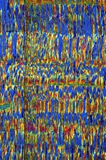
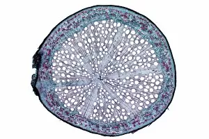
Beech tree stem. Polarised light micrograph of a section through the woody stem of a common beech tree (Fagus sylvatica), showing diffuse-porous xylem vessels (dark blue)

Background imageImages Dated 18th January 2012: Geranium stem, light micrograph

Geranium stem, light micrograph
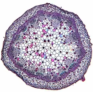
Geranium stem. Light micrograph of a section through a young stem of a geranium (Pelargonium sp.) plant. The outer stem is covered with a thin epidermis (red) which has stomata

Background imageImages Dated 18th January 2012: Edme Pajot, French physician

Edme Pajot, French physician
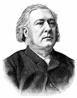
Charles Marie Edme Pajot (1816-1896), French physician. Pajot was an obstetrician and gynaecologist. He studied and worked in Paris

Background imageImages Dated 18th January 2012: Sweet pea stem, light micrograph

Sweet pea stem, light micrograph
Sweet pea stem. Light micrograph of a section through the hollow stem of a sweet pea (Lathyrus odoratus) plant, showing a ring of vascular bundles

Background imageImages Dated 18th January 2012: Railroad drawbridge, 19th century

Railroad drawbridge, 19th century
Railroad drawbridge, 19th-century artwork. This type of bridge is known as a bascule bridge. This one is near Rutherford, New Jersey, USA, on the Erie Railroad Main Line

Background imageImages Dated 18th January 2012: Orange fruit, light micrograph

Orange fruit, light micrograph
Orange fruit. Light micrograph of a section through the rind of a fruit from a bitter orange (Citrus aurantium) tree, showing large numbers of spherical oil-secreting glands

Background imageImages Dated 18th January 2012: Shepherds purse fruit, light micrograph

Shepherds purse fruit, light micrograph
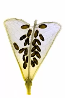
Shepherds purse fruit. Light micrograph of a section through a seed pod from a shepherds purse (Capsella bursa-pastoris) plant. The remains of the stigma can be seen (top centre)

Background imageImages Dated 18th January 2012: Sage stem, light micrograph

Sage stem, light micrograph
Sage stem. Light micrograph of a section through a primary stem of a scarlet sage (Salivia splendens) plant. The outer stem is covered with a thin epidermis (green) that contains stomata

Background imageImages Dated 18th January 2012: Rhubarb root, light micrograph

Rhubarb root, light micrograph
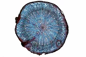
Rhubarb root. Light micrograph of a section through the root of a rhubarb (Rheum sp.) plant. The primary cortex has been shed by the formation of a circular meristem, the periderm

Background imageImages Dated 18th January 2012: August Kekule, German chemist

August Kekule, German chemist
August Kekule (1829-1896), German organic chemist. From 1858 Kekule developed a structural theory for carbon chemistry, based on its valency (bond-forming) of four

Background imageImages Dated 18th January 2012: Sycamore leaf in autumn, micrograph

Sycamore leaf in autumn, micrograph
Sycamore leaf in autumn. Light micrograph of a section through the stem of a leaf from a sycamore (Acer pseudoplantanus) tree in autumn

Background imageImages Dated 18th January 2012: Beech tree leaf, light micrograph

Beech tree leaf, light micrograph
Beech tree leaf. Light micrograph of a section through the leaf of a common beech tree (Fagus sylvatica), showing the midrib

Background imageImages Dated 18th January 2012: Lime tree stem wound, light micrograph

Lime tree stem wound, light micrograph
Lime tree stem wound. Light micrograph of a section through the wounded stem of a lime tree (Tilia europaea). The outer epidermis has been shed and replaced by a layer of cork (dark red)

Background imageImages Dated 18th January 2012: Marrow stem, light micrograph

Marrow stem, light micrograph
Marrow stem. Light micrograph of a section through the stem of a marrow (Curcurbita sp.), showing the sieve plates in the phloem. A single collateral vascular bundle can be seen

Background imageImages Dated 18th January 2012: Common sundew, light micrograph

Common sundew, light micrograph
Common sundew. Light micrograph of a section through the leaf of a common sundew (Drosera rotundifolia) plant, showing the glandular hairs (dark)

Background imageImages Dated 18th January 2012: Sage stem, light micrograph

Sage stem, light micrograph
Sage stem. Light micrograph of a section through a secondary stem of a scarlet sage (Salivia splendens) plant. The outer stem is covered with a thin epidermis and thick cuticle (red)

Background imageImages Dated 18th January 2012: Oak root, light micrograph

Oak root, light micrograph
Oak root. Light micrograph of a section through a secondary root from an oak (Quercus sp.) tree. The primary cortex (outer layer) has been shed by the formation of a circular meristem, the periderm

Background imageImages Dated 18th January 2012: Dwarfism, 19th century

Dwarfism, 19th century
Dwarfism, 19th-century artwork. Burmese dwarfs Smaun (left) and Fatma (right) standing next to a champagne bottle. These two dwarfs, brother (Smaun) and sister (Fatma), had the surname Sing Hpoo

Background imageImages Dated 18th January 2012: Magic trick, 19th century

Magic trick, 19th century artwork. Shown here is the double cage magic trick, where the magician makes one or both of his assistants (in the cages) disappear or swap places

Background imageImages Dated 18th January 2012: Ancient Egyptian artefact, 19th century

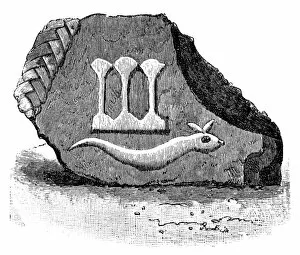
Ancient Egyptian artefact, 19th century artwork. This artefact, found in Egypt in the 1890s, is a fragment of an Ancient Egyptian object called a pschent

Background imageImages Dated 18th January 2012: Lupin root nodules, light micrograph

Lupin root nodules, light micrograph
Lupin root nodules. Light micrograph of a section through the root nodules of a lupin (Lupinus lupin) plant. These nodules are full of nitrogen-fixing Rhizobium sp

Background imageImages Dated 18th January 2012: Strongwoman, 19th century

Strongwoman, 19th century
Strongwoman, 19th-century artwork. Strongwoman Mary Arniotis lifting five men in a performance in Berlin, Germany. Arniotis performed feats of strength while touring Europe and the USA in the 1890s

Background imageImages Dated 18th January 2012: African mahogany stem, light micrograph

African mahogany stem, light micrograph
African mahogany stem. Polarised light micrograph of a section through the stem of an African mahogany (Khaya sp.) tree. The multicoloured cells are fibres, and fibre tracheids, which carry water

Background imageImages Dated 18th January 2012: Shepherds purse fruit, light micrograph

Shepherds purse fruit, light micrograph
Shepherds purse fruit. Light micrograph of a section through a seed pod from a shepherds purse (Capsella bursa-pastoris) plant. The remains of the stigma can be seen (top centre)

Background imageImages Dated 18th January 2012: White bryony stem, light micrograph

White bryony stem, light micrograph
White bryony stem. Light micrograph of a transverse section through the stem of a white bryony (Bryonia alba) plant, showing a single collateral vascular bundle

Background imageImages Dated 18th January 2012: Beech tree stem, light micrograph

Beech tree stem, light micrograph
Beech tree stem. Light micrograph of a section through the woody stem of a common beech tree (Fagus sylvatica). From left to right: oval multiseriate ray (orange)

Background imageImages Dated 18th January 2012: Lime tree stem, light micrograph

Lime tree stem, light micrograph
Lime tree stem. Light micrograph of a section through the stem of a lime tree (Tilia europaea). The outer epidermis has been shed and replaced by a layer of cork (dark red)

Background imageImages Dated 18th January 2012: Beech tree leaves, light micrograph

Beech tree leaves, light micrograph
Beech tree leaves. Light micrograph of a section through two leaves from different parts of a common beech tree (Fagus sylvatica)

Background imageImages Dated 18th January 2012: Pedal-powered vessel, 19th century

Pedal-powered vessel, 19th century
Pedal-powered vessel, 19th-century artwork. This vessel has two helical screws either side, powered by pedals operated by four cyclists, with a fifth seat at the front for the pilot

Background imageImages Dated 18th January 2012: Root vegetable washer, 19th century

Root vegetable washer, 19th century artwork. This invention was designed by a Mr Defosse-Delambre, from Varennes, in the Somme region of France

Background imageImages Dated 18th January 2012: Orange fruit, light micrograph

Orange fruit, light micrograph
Orange fruit. Light micrograph of a section through the rind of a fruit from a bitter orange (Citrus aurantium) tree, showing one of the many spherical oil-secreting glands

Background imageImages Dated 18th January 2012: African mahogany stem, light micrograph

African mahogany stem, light micrograph
African mahogany stem. Polarised light micrograph of a section through the stem of an African mahogany (Khaya sp.) tree. The multicoloured cells are fibres, and fibre tracheids, which carry water

Background imageImages Dated 18th January 2012: Poppy flower bud, light micrograph

Poppy flower bud, light micrograph
Poppy flower bud. Light micrograph of a section through a flower bud from a poppy (Papaver sp.) plant. From outside to inside: calyx, made up of the sepals; corolla

Background imageImages Dated 18th January 2012: Venus flytrap leaf, light micrograph

Venus flytrap leaf, light micrograph
Venus flytrap leaf. Light micrograph of a section through the leaf of a Venus flytrap (Dionaea sp.), showing the digestive glands that line its interior

Background imageImages Dated 18th January 2012: African teak stem, light micrograph

African teak stem, light micrograph
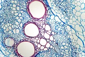
African teak stem. Light micrograph of a section through the woody stem of an African teak (Pericopsis elata) plant, showing its diffuse-porous xylem tissue

Background imageImages Dated 18th January 2012: Nansens Fram expedition, 19th century

Nansens Fram expedition, 19th century
Fram expedition to the Arctic, commanded by the Norwegian explorer Fridtjof Nansen (1861-1930). Nansen and his crew sailed in the Fram (top left)

Background imageImages Dated 18th January 2012: Gunboat on Nile rapids, 19th century

Gunboat on Nile rapids, 19th century
Gunboat on Nile rapids, 19th-century artwork. This is a British gunboat being handled by ropes as it negotiates rapids or cataracts on the Nile

Background imageImages Dated 18th January 2012: Oleander leaf, light micrograph

Oleander leaf, light micrograph
Oleander leaf. Light micrograph of a section through the leaf of an oleander (Nerium oleander) tree, showing its sunken stomata (gaps, centre left and right)


All Professionally Made to Order for Quick Shipping


-

Why Choose Us?

We are a leading provider of Art Prints and Photo Gifts since 2003, working in partnership with a range of Sporting Clubs, Charities, Museums and Picture Libraries. A large share of profits from any of their images will go directly towards supporting that charity or club. Our archive of images is carefully curated to bring you a wide range of subjects, including landscapes, wildlife, architecture, and more. We ship from our partner labs in the UK, USA, EU (Netherlands) and Australia.
+

How do I place an order?

Ordering is quick & easy - Just follow 5 Simple Steps:
  1. Find your image: Use our search box or browse our online photo Collections to find the image you want.

  2. Choose your print format: Select your desired product and add it to your cart.

  3. Enter your details: If you're a returning customer, simply enter your email address and password, and we'll fill in your billing and shipping address details. All personal details are held securely and are fully GDPR compliant. As standard, we remove all Personally Identifiable Information after 12 months.

  4. Pay for your purchase: We use state-of-the-art security for online shopping and do not have access to your card details.

  5. Sit back and relax: We'll email you confirmation of your order and when it's dispatched. Registered customers can also track orders in the 'My Account' area.

+

How do I pay for an order?

You can pay for your order with most of the major credit and debit cards, or PayPal. For added security, major financial institutions process payment details separately and securely on our behalf. We do not have access to your online payment card details. Online payments are preferred however we do take phone orders. For UK customers only we accept cheques issued against a UK bank.
+

Is my data safe?

We take data security very seriously. We do not have access to your full card details and all payments are requested over a fully secure connection. Additionally, we fully comply with current European and GDPR legislation, and automatically remove all personal data after at most 12 months (unless you have an account with us and have signed in recently). We also have a strict opt-in policy and would never sell your personal details. Your data is only used to fulfill your order promptly and efficiently.
+

Is wall art ready to hang?

For quick and easy installation all wall art, including framed prints, canvas prints and metal prints are supplied with a ready to hang solution on the back. Generally, saw tooth hangers are applied as they allow wall art to hang flush against the wall. The serrated edge of the hanger prevents the frame from shifting or tilting when hung.
+

Are the photo prints fade resistant?

Yes, we use archival quality photo paper photographic paper for vivid reproduction Prints are an accurate representations of the original artwork, which is preserved for artistic character and authenticity. We guarantee they match previews shown on our web site