Fraud Blocker Skip to main content

Home > Science > SEM
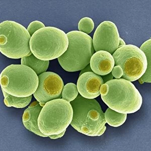
SEM of yeast cells

 : SEM of yeast cells
In Stock
We accept all major payment options so you can pay for the things you need in the way that is most convenient for you

Choose Your Product

Framed Photos
Buy from AU$119.99
Photo Prints
Buy from AU$12.99
Jigsaw Puzzles
Buy from AU$89.99
Poster Prints
Buy from AU$24.99
Canvas Prints
Buy from AU$124.99
Fine Art Prints
Buy from AU$94.05
Metal Prints
Buy from AU$43.99
Cards
Buy from AU$12.99
Home Decor
Buy from AU$24.99

Science Photo Library Photo Prints and Wall Art

SEM of yeast cells

Yeast. Coloured scanning electron micrograph (SEM) of yeast cells, Saccharomyces cerevisiae, commonly known as Bakers or Brewers yeast, growing on potato dextrose agar. Yeast is able to ferment sugar, producing alcohol & carbon dioxide in the process. It has long been used in the brewing of beer, the production of wine, and in baking of leavened bread (carbon dioxide causes the dough to rise). Medically, dried yeast is used as a source of vitamin B1, riboflavin & nicotinic acid. Yeast divide rapidly by budding off new cells (visible here). They may also reproduce sexually. Magnification: X945 at 35mm size. Coloured orange. Original is BW print B250/174. 12 -- damaged. Science Photo Library features Science and Medical images including photos and illustrations. © DR JEREMY BURGESS/SCIENCE PHOTO LIBRARY

Media ID 6292169

Budding Eumycota Fungal Fungi Fungus Mycology Naturemycology Saccharomyces Cerevisiae Yeast

FEATURES IN THESE COLLECTIONS

> Arts > Artists > Z > Zucchero

> Science > SEM

SECURE PAYMENTS
We accept all major payment options so you can pay for the things you need in the way that is most convenient for you

Made in Australia
Safe Shipping, Easy Returns

FREE PERSONALISATION
We are proud to offer a range of customisation including Cropping, Captions, and Filters. Zoom Cropping can be adjusted in the basket


EDITORS COMMENTS

This print showcases a coloured scanning electron micrograph (SEM) of yeast cells, specifically Saccharomyces cerevisiae, also known as Bakers or Brewers yeast. The image captures the yeast cells growing on potato dextrose agar, highlighting their intricate structures and vibrant orange hue. Yeast is renowned for its ability to ferment sugar, producing both alcohol and carbon dioxide in the process. This remarkable quality has made it an essential ingredient in brewing beer, producing wine, and leavening bread dough by causing it to rise through the release of carbon dioxide. Beyond culinary applications, dried yeast serves as a valuable source of vitamin B1, riboflavin, and nicotinic acid in medical contexts. In this SEM image, we can observe how yeast cells rapidly divide by budding off new cells—a fascinating sight that emphasizes their reproductive capabilities. The magnification level of X945 at 35mm size allows us to appreciate the intricate details of these tiny organisms. While this particular print suffered some damage during its journey through time, it still manages to captivate viewers with its scientific beauty. Overall, this photograph offers a glimpse into the microscopic world of fungi—showcasing nature's incredible diversity and reminding us of the significant role that yeasts play in various fields such as food production and medicine.


Framed Prints of SEM of yeast cells

Discover the fascinating world of microbiology with Media Storehouse's Framed Prints featuring this captivating Coloured Scanning Electron Micrograph (SEM) of yeast cells by Science Photo Library. Witness the intricate detail of Saccharomyces cerevisiae, also known as Bakers or Brewers yeast, as it grows on potato dextrose agar. This high-quality framed print brings the beauty of science right into your home or office, making it an excellent addition for any space inspired by biology, microbiology, or the wonders of nature.


Photo Prints of SEM of yeast cells

Discover the intricacies of the microscopic world with our Media Storehouse range of Photographic Prints. This captivating image showcases a Coloured Scanning Electron Micrograph (SEM) of Saccharomyces cerevisiae, also known as Bakers or Brewers yeast, growing on potato dextrose agar. Witness the mesmerizing details of these yeast cells, adding a touch of science and beauty to your space. Our high-quality prints preserve the vibrant colours and fine details of this Science Photo Library photograph, making it an excellent addition to any laboratory, classroom, or home decor. Explore the wonders of the universe with Media Storehouse.


Poster Prints of SEM of yeast cells

Discover the intricacy of the microscopic world with Media Storehouse's Poster Prints featuring this captivating Coloured Scanning Electron Micrograph (SEM) of Saccharomyces cerevisiae, or commonly known as Bakers or Brewers yeast, growing on potato dextrose agar. This stunning image, sourced from Science Photo Library, showcases the mesmerizing detail of yeast cells in their natural environment. Ideal for laboratories, classrooms, or any space that appreciates the beauty of science, our high-quality poster prints bring a touch of exploration and discovery to your walls.


Jigsaw Puzzles of SEM of yeast cells

Discover the fascinating world of microbiology with our Media Storehouse Jigsaw Puzzles! This intriguing puzzle, featuring a Coloured Scanning Electron Micrograph (SEM) of Saccharomyces cerevisiae, or yeast cells, by Science Photo Library, offers a unique and educational experience. Delve into the complex structures of these essential organisms, commonly used in baking and brewing industries. Assemble the puzzle pieces to reveal the stunning detail and vibrant colors of the yeast cells growing on potato dextrose agar. A perfect activity for science enthusiasts, families, or anyone looking for a captivating and thought-provoking puzzle experience.

diamond
Made With High Grade Materials
thumb_up
Professional Quality Finish
package_2
Carefully Packed To Aid Safe Arrival

You May Also Like


Wood. Scanning electron microscope (SEM)Wood. Scanning electron microscope (SEM)
Add to Basket
Yeast cells, SEMYeast cells, SEM
Add to Basket
Oldham High Street 1949Oldham High Street 1949
Add to Basket
Gordon Solomon playing the trombone in the Newcastle Big Band in February 1973Gordon Solomon playing the trombone in the Newcastle Big Band in February 1973
Add to Basket
Children: Clever: Master Marvel: A mini mastermind is to star in a television documentaryChildren: Clever: Master Marvel: A mini mastermind is to star in a television documentary
Add to Basket
The Sahara Camel - Special Breed - youngsters have black nosesThe Sahara Camel - Special Breed - youngsters have black noses
Add to Basket
PIC SHOWS SWIMWEAR FASHION WITH MODEL JULIE HANNAHPIC SHOWS SWIMWEAR FASHION WITH MODEL JULIE HANNAH
Add to Basket
Keeping in touch with home. This simple post box has been made out of a dis-usedKeeping in touch with home. This simple post box has been made out of a dis-used
Add to Basket
Sheep in the salt marsh nature reserve, Hohwacht Bay, Behrensdorf, Baltic Sea, Schleswig-Holstein, Germany, EuropeSheep in the salt marsh nature reserve, Hohwacht Bay, Behrensdorf, Baltic Sea, Schleswig-Holstein, Germany, Europe
Add to Basket
To erect a monumentTo erect a monument
Add to Basket
Godfrey of Bouillon (engraving)Godfrey of Bouillon (engraving)
Add to Basket
Aerail view at Ehrenburg castle, Brodenbach, Mosel valley, Hunsruck, Rhineland-Palatinate, GermanyAerail view at Ehrenburg castle, Brodenbach, Mosel valley, Hunsruck, Rhineland-Palatinate, Germany
Add to Basket

Gallery Wall Inspiration

Nerve cell, SEM
Red-barbed ant, SEM
Neutrophil engulfing MRSA, SEM C018 / 8596
Diatom alga, SEM
Diatom, SEM
X and Y chromosomes
Lavender pollen grain, SEM
Scanning Electron Micrograph (SEM): Tardigrade or ‘Water Bear Magnification x 1250 (A4 size: 29. 7 cm width)
Cat tongue surface, SEM
False-coloured SEM (Scanning Electron Micrograph) of a Tardigrade (Tardigrada) amongst moss, from Peak District National Park, Derbyshire, UK. This tardigrade is approximately 0. 1 millimetres long. Digital composite
Hazel pollen grains
Collagen, Scanning electron micrograph (SEM)
Santos-Dumont / Sem
T lymphocytes and cancer cell, SEM C001 / 1679
Tardigrade or Water Bear
Crysotile asbestos
Uterus lining during menstruation, SEM
Iris of the eye, SEM
Stylus in LP record
Salmonella bacteria, SEM
SEM of penicillin fungus
Vascular bundle, SEM
Geranium anther, SEM
Dahlia flower pollen, SEM
Scanning Electron Micrograph (SEM): Praying Mantis - Magnification x 30 (if print A4 size: 29. 7 cm wide)
Tardigrade, SEM
Scanning electron micrograph (SEM) of red blood cell


© Copyright Media Storehouse, All Rights Reserved 2003 - 2025
Facebook     Pinterest     100% Payment Secure     Reviews IO