Fraud Blocker Skip to main content

Home > Science > SEM

SEM Collection (page 8)

1,336 items

Background imageSEM Collection: Lemon grass, SEM

Lemon grass, SEM
Lemon grass. Coloured scanning electron micrograph (SEM) of the surface of Thai lemon grass (Cymbopogon flexuosus) showing closed stomata holes

Background imageSEM Collection: Mitochondria, SEM

Mitochondria, SEM
Mitochondria. Coloured scanning electron micrograph (SEM) of mitochondria (green) in a kidney cell. Mitochondria are a type of organelle found in the cytoplasm of eukaryotic cells

Background imageSEM Collection: Vanilla pod, SEM

Vanilla pod, SEM
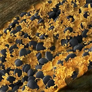
Vanilla pod, coloured scanning electron micrograph (SEM). Section through a vanilla (Vanilla planifolia) pod showing the black seeds and parachyma tissue

Background imageSEM Collection: Chickweed pollen grains, SEM

Chickweed pollen grains, SEM
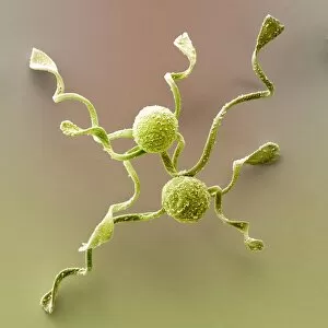
Chickweed pollen grains. Coloured scanning electron micrograph (SEM) of pollen grains (green) from a chickweed flower (Stellaria media)

Background imageSEM Collection: Diatoms, SEM

Diatoms, SEM
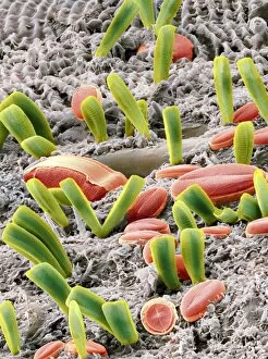
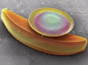
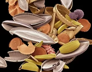
Diatoms. Coloured scanning electron micrograph (SEM) of various freshwater diatoms attached to algae (grey). The diatoms are a group of photosynthetic, single-celled algae containing about 100

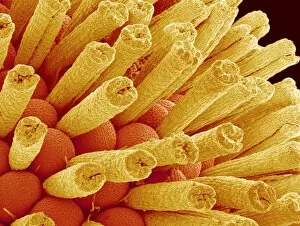
Background imageSEM Collection: Ladys mantle reproductive parts, SEM

Ladys mantle reproductive parts, SEM
Ladys mantle reproductive parts, coloured scanning electron micrograph (SEM). Ladys mantle, a plant in the genus Alchemilla

Background imageSEM Collection: Macrophage engulfing TB bacteria, SEM

Macrophage engulfing TB bacteria, SEM
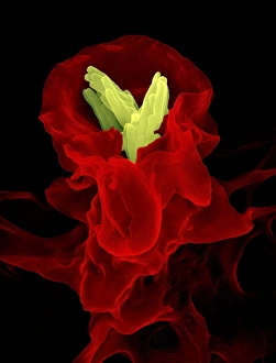
Macrophage engulfing TB bacteria. Coloured scanning electron micrograph (SEM) of a macrophage white blood cell (red) engulfing a tuberculosis (Mycobacterium tuberculosis) bacterium (yellow)

Background imageSEM Collection: Bread mould, SEM

Bread mould, SEM
Bread mould. Coloured scanning electron micrograph (SEM) of a sporangium of a bread mould (Rhizopus stolonifer). Sporangia are asexual reproductive structures where the moulds spores develop before

Background imageSEM Collection: Moon rock, SEM

Moon rock, SEM
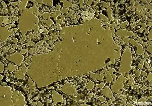
Moon rock. Coloured scanning electron micrograph (SEM) of the surface of a sample of rock from the Moon

Background imageSEM Collection: Splenda artificial sweetener, SEM

Splenda artificial sweetener, SEM
Splenda artificial sweetener. Coloured scanning electron micrograph (SEM) of crystals of Splenda. Splenda is an artificial sweetener made from dextrose and Sucralose

Background imageSEM Collection: Tick mouthparts, SEM

Tick mouthparts, SEM
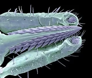
Tick mouthparts, coloured scanning electron micrograph (SEM). The mouthparts of a tick consist of three visible components. The two outer jointed parts are highly mobile palps

Background imageSEM Collection: Euphorbia flower reproductive parts, SEM

Euphorbia flower reproductive parts, SEM
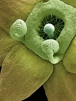
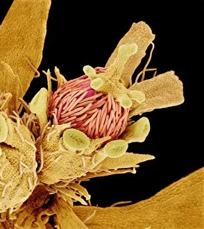
Euphorbia flower reproductive parts. Coloured scanning electron micrograph (SEM) of the reproductive parts of a Euphorbia supina flower

Background imageSEM Collection: Bacteria infecting a macrophage, SEM

Bacteria infecting a macrophage, SEM
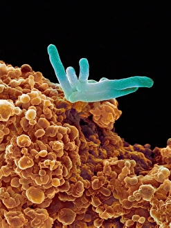
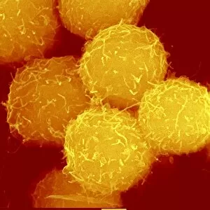
Bacteria infecting a macrophage. Coloured scanning electron micrograph (SEM) of Mycobacterium tuberculosis bacteria (green) infecting a macrophage white blood cell

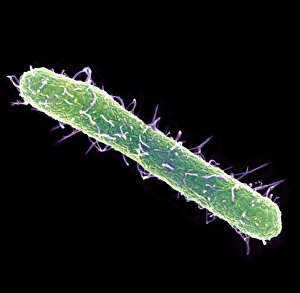
Background imageSEM Collection: Salmonella typhimurium bacterium, SEM

Salmonella typhimurium bacterium, SEM
Salmonella typhimurium bacterium, coloured scanning electron micrograph (SEM). This bacterium is also known as Salmonella enterica serovar typhimurium

Background imageSEM Collection: Diatom alga, SEM

Diatom alga, SEM
Diatom. Coloured scanning electron micrograph (SEM) of the surface of the mineralised cell wall (frustule) of an unidentified diatom. This is a planktonic unicellular alga

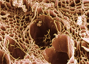
Background imageSEM Collection: Rotten wood, SEM

Rotten wood, SEM
Rotten wood. Coloured scanning electron micrograph (SEM) of a piece of domestic plywood infected with dry rot fungus. The structure of the wood is seen here

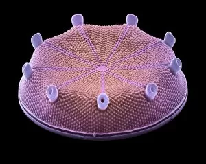
Background imageSEM Collection: Diatom alga, SEM

Diatom alga, SEM
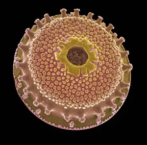
Diatom. Coloured scanning electron micrograph (SEM) of an Aulacodiscus oregonus diatom. This is a marine planktonic unicellular alga

Background imageSEM Collection: Geranium pollen, SEM

Geranium pollen, SEM
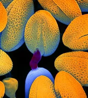
Geranium pollen. Coloured scanning electron micrograph (SEM) of pollen (orange) on the anther (yellow) of a geranium flower (Geranium sp.)

Background imageSEM Collection: Wild strawberry flower pistil, SEM

Wild strawberry flower pistil, SEM
Wild strawberry pistil. Coloured scanning electron micrograph (SEM) of the pistil of a flower of the Asian wild strawberry (Potentilla sp.). The pistil is the female reproductive part of a flower

Background imageSEM Collection: Geranium pollen, SEM

Geranium pollen, SEM
Geranium pollen. Coloured scanning electron micrograph (SEM) of pollen (pink) on the stamen of a geranium flower (Geranium sp.)

Background imageSEM Collection: Pussy willow pollen

Pussy willow pollen
Willow pollen. Coloured scanning electron micro- graph (SEM) of a pollen grain of goat willow, Salix caprea. Also known as the " pussy willow"

Background imageSEM Collection: Germination of turnip pollen

Germination of turnip pollen
Germinating pollen grain. Coloured scanning electron micrograph (SEM) showing the germination of a pollen grain on the stigma of a turnip flower, Brassica campestris

Background imageSEM Collection: Orchid petal, SEM

Orchid petal, SEM
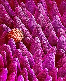
Orchid petal. Coloured scanning electron micrograph (SEM) of the surface of an orchid petal (Cymbidium sp. ). It is covered in tiny epidermal hairs, or trichomes

Background imageSEM Collection: French lavender leaf surface, SEM

French lavender leaf surface, SEM
French lavender leaf surface. Coloured scanning electron micrograph (SEM) of a section of French lavender (Lavandula dentata) leaf, showing the tooth-like structures (trichomes)

Background imageSEM Collection: Olive leaf trichomes, SEM

Olive leaf trichomes, SEM
Olive leaf trichomes. Coloured scanning electron micrograph (SEM) of peltate trichomes, or modified hairs, on the surface of an olive leaf (Olea europaea)

Background imageSEM Collection: Leaf oil glands, SEM

Leaf oil glands, SEM
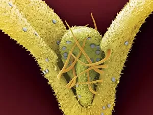
Leaf oil glands. Coloured scanning electron micrograph (SEM) of leaves from a thyme plant (Thymus vulgaris). A flower bud is seen between the two leaves. Two types of leaf trichomes (hairs) are seen

Background imageSEM Collection: Primrose pollen, SEM

Primrose pollen, SEM
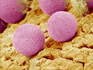
Primrose pollen. Coloured scanning electron micrograph (SEM) of pollen grains (pink) from a primrose flower (Primula vulgaris)

Background imageSEM Collection: Flowers, SEM

Flowers, SEM
Flowers. Coloured scanning electron micrograph (SEM) of unidentified wild flowers. Magnification unknown

Background imageSEM Collection: Sunflower pollination, SEM

Sunflower pollination, SEM
Sunflower pollination. Coloured scanning electron micrograph (SEM) of pollen on a stigma of a sunflower plant (Helianthus sp.)

Background imageSEM Collection: Pansy petal, SEM

Pansy petal, SEM
Pansy petal. Coloured scanning electron micrograph (SEM) of the surface of a pansy flower petal (Viola tricolor). The petal is covered in tiny epidermal hairs known as trichomes

Background imageSEM Collection: Primrose stigma, SEM

Primrose stigma, SEM
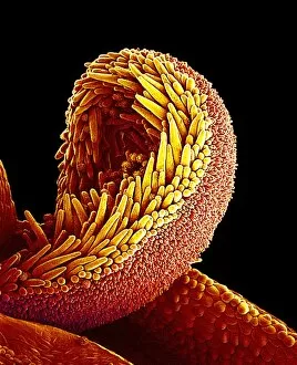
Primrose stigma. Coloured scanning electron micrograph (SEM) of the stigma (yellow) and style (lower left) of a primrose flower (Primula sp.)

Background imageSEM Collection: Bacteria in the nose, SEM

Bacteria in the nose, SEM
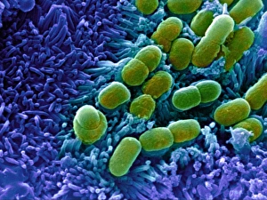
Bacteria in the nose. Coloured scanning electron micrograph (SEM) of bacteria (orange) on the surface of the nasal cavity

Background imageSEM Collection: E. coli bacteria, SEM

E. coli bacteria, SEM
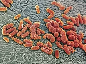
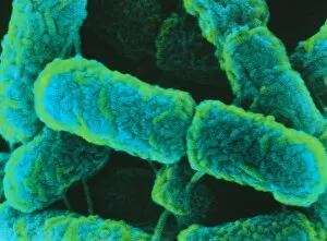
E. coli bacteria. Coloured scanning electron micrograph (SEM) of Escherichia coli bacteria. These bacilli (rod-shaped bacteria)

Background imageSEM Collection: Fallopian tube cells, SEM

Fallopian tube cells, SEM
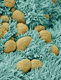
Brain lining. Coloured scanning electron micrograph (SEM) of the lining of the brain, showing ependymal cells (yellow) and ciliary hairs (green)

Background imageSEM Collection: Diatom, SEM

Diatom, SEM
Diatom. Coloured scanning electron micrograph (SEM) of a Triceratium sp. diatom. Diatoms are single-celled photosynthetic algae, of which there are about 100, 000 species

Background imageSEM Collection: Diatoms, SEM

Diatoms, SEM
Diatoms, coloured scanning electron micrograph (SEM). These are marine planktonic unicellular algae. They have a mineralised cell wall (frustule)

Background imageSEM Collection: Diatom, SEM

Diatom, SEM
Diatom. Coloured scanning electron micrograph (SEM) of a diatom. Diatoms are single-celled photosynthetic algae, of which there are about 100

Background imageSEM Collection: Diatoms, SEM

Diatoms, SEM
Diatoms. Coloured scanning electron micrograph (SEM) of diatoms. The diatoms are a group of photosynthetic, single-celled algae containing about 10, 000 species

Background imageSEM Collection: Diatom, SEM

Diatom, SEM
Diatom. Coloured scanning electron micrograph (SEM) of a Stephanopyxis sp. diatom. Diatoms are single-celled photosynthetic algae, of which there are about 100, 000 species

Background imageSEM Collection: Coloured SEM of Escherichia coli bacteria

Coloured SEM of Escherichia coli bacteria
E. coli bacteria. Coloured scanning electron micrograph of the rod-shaped, Gram-negative bacteria, Escherichia coli, commonly known as E. coli

Background imageSEM Collection: E. coli bacteria, SEM

E. coli bacteria, SEM
E. coli bacteria. Coloured scanning electron micrograph (SEM) of Escherichia coli bacteria (green) taken from the small intestine of a child. E

Background imageSEM Collection: Diatom, SEM

Diatom, SEM
Diatom. Coloured scanning electron micrograph (SEM)of the diatom Coscinodiscus sp. The diatoms are a group of photosynthetic, single-celled algae containing about 10, 000 species

Background imageSEM Collection: Fungal spores, SEM

Fungal spores, SEM
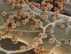
Fungal spores. Coloured scanning electron micrograph (SEM) of the spores of an unidentified fungus (red) on a cats hair

Background imageSEM Collection: Horsetail spores, SEM

Horsetail spores, SEM
Horsetail spores. Coloured scanning electron micrograph (SEM) of spores of a horsetail (Equisetum arvense) plant. The spores (round) bear elaters (helical)

Background imageSEM Collection: Scanning Electron Micrograph (SEM): Follicle Mite - Magnification x 3800 (if print A4 size: 29)

Scanning Electron Micrograph (SEM): Follicle Mite - Magnification x 3800 (if print A4 size: 29. 7 cm wide)
LRDS-424 Scanning Electron Micrograph (SEM): Follicle Mite Magnification x 3800 (if print A4 size: 29.7 cm wide) Demodex folliculorum David Spears (Last Refuge)

Background imageSEM Collection: Scanning Electron Micrograph (SEM): Common Clothes Moth - Magnification x 35 (if print A4 size)

Scanning Electron Micrograph (SEM): Common Clothes Moth - Magnification x 35 (if print A4 size: 29. 7 cm wide)
LRDS-401 Scanning Electron Micrograph (SEM): Common Clothes Moth Magnification x 35 (if print A4 size: 29.7 cm wide) Tineola bisselliella David Spears (Last Refuge)

Background imageSEM Collection: Scanning Electron Micrograph (SEM): Common Rough Woodlouse, Magnification x 20 (A4 size: 29)

Scanning Electron Micrograph (SEM): Common Rough Woodlouse, Magnification x 20 (A4 size: 29. 7 cm width)
LRDS-148 Common Rough Woodlouse Scanning Electron Micrograph (SEM) Porcellio scaber Magnification x 20 (A4 size: 29.7 cm width) Coloured by hand to enhance natural features

Background imageSEM Collection: Haematopoietic stem cells, SEM C013 / 5009

Haematopoietic stem cells, SEM C013 / 5009
Haematopoietic stem cells, coloured scanning electron micrograph (SEM). Stem cells can differentiate into any other cell type


All Professionally Made to Order for Quick Shipping


-

Why Choose Us?

We are a leading provider of Art Prints and Photo Gifts since 2003, working in partnership with a range of Sporting Clubs, Charities, Museums and Picture Libraries. A large share of profits from any of their images will go directly towards supporting that charity or club. Our archive of images is carefully curated to bring you a wide range of subjects, including landscapes, wildlife, architecture, and more. We ship from our partner labs in the UK, USA, EU (Netherlands) and Australia.
+

How do I place an order?

Ordering is quick & easy - Just follow 5 Simple Steps:
  1. Find your image: Use our search box or browse our online photo Collections to find the image you want.

  2. Choose your print format: Select your desired product and add it to your cart.

  3. Enter your details: If you're a returning customer, simply enter your email address and password, and we'll fill in your billing and shipping address details. All personal details are held securely and are fully GDPR compliant. As standard, we remove all Personally Identifiable Information after 12 months.

  4. Pay for your purchase: We use state-of-the-art security for online shopping and do not have access to your card details.

  5. Sit back and relax: We'll email you confirmation of your order and when it's dispatched. Registered customers can also track orders in the 'My Account' area.

+

How do I pay for an order?

You can pay for your order with most of the major credit and debit cards, or PayPal. For added security, major financial institutions process payment details separately and securely on our behalf. We do not have access to your online payment card details. Online payments are preferred however we do take phone orders. For UK customers only we accept cheques issued against a UK bank.
+

Is my data safe?

We take data security very seriously. We do not have access to your full card details and all payments are requested over a fully secure connection. Additionally, we fully comply with current European and GDPR legislation, and automatically remove all personal data after at most 12 months (unless you have an account with us and have signed in recently). We also have a strict opt-in policy and would never sell your personal details. Your data is only used to fulfill your order promptly and efficiently.
+

Is wall art ready to hang?

For quick and easy installation all wall art, including framed prints, canvas prints and metal prints are supplied with a ready to hang solution on the back. Generally, saw tooth hangers are applied as they allow wall art to hang flush against the wall. The serrated edge of the hanger prevents the frame from shifting or tilting when hung.
+

Are the photo prints fade resistant?

Yes, we use archival quality photo paper photographic paper for vivid reproduction Prints are an accurate representations of the original artwork, which is preserved for artistic character and authenticity. We guarantee they match previews shown on our web site