Xray Collection
X-rays are a type of electromagnetic radiation that have shorter wavelengths than visible light
Choose a picture from our Xray Collection for your Wall Art and Photo Gifts
1,307 items
All Professionally Made to Order for Quick Shipping
-
Xray Collection
X-rays are a type of electromagnetic radiation that have shorter wavelengths than visible light. They were discovered in 1895 by German physicist Wilhelm Conrad Röntgen, who found that they could penetrate through solid objects and produce images on photographic plates. X-rays are used extensively in medical imaging to diagnose and treat various conditions, such as broken bones, tumors, and dental problems and can also be used for non-medical purposes, such as studying the structure of materials or detecting hidden defects in manufactured products. However, exposure to high levels of X-rays can be harmful to human health due to their ionizing nature.
+
Our beautiful pictures are available as Framed Prints, Photos, Wall Art and Photo Gifts
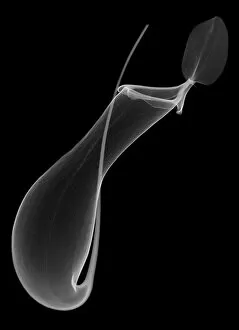
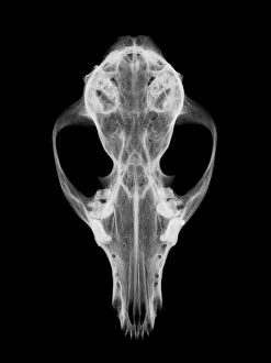
The Xray collection from Media Storehouse offers a unique and fascinating glimpse into the world of science. Our collection features stunning images of various objects, animals, and even humans captured using x-ray technology. The intricate details and inner workings of these subjects are revealed in a way that is both beautiful and informative. From the delicate bones of birds to the complex machinery inside industrial equipment, our collection showcases the incredible versatility of x-ray imaging. These prints make for striking wall art or framed pieces that will add an element of curiosity to any room. Whether you're a science enthusiast or simply appreciate visually stunning artwork, the Xray collection from Media Storehouse is sure to impress. With high-quality prints available in a range of sizes, there's something for everyone in this captivating collection.
+
What are Xray (Science) art prints?
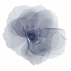
Xray art prints are a unique and fascinating collection of images that showcase the beauty and complexity of the natural world. These prints feature X-ray photographs of various objects, from flowers to animals, revealing their intricate inner workings in stunning detail. The use of X-rays allows us to see beyond what is visible to the naked eye, providing a new perspective on familiar subjects. These art prints are perfect for those who appreciate science and nature, as well as anyone looking for something truly unique to decorate their home or office. They make great conversation starters and can be used to add interest and depth to any space. We offer a wide range of Xray art prints in different sizes and formats, including framed options for easy display. Each print is produced using high-quality materials to ensure long-lasting durability and vibrant colors that will stand out wherever they are displayed.
+
What Xray (Science) art prints can I buy from Media Storehouse?
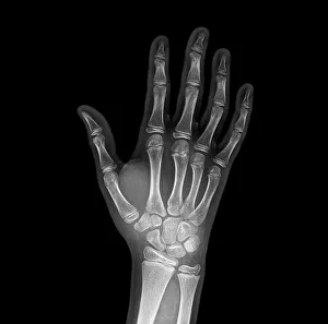
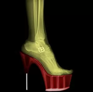
At Media Storehouse, you can find a wide range of Xray art prints that are perfect for adding a unique touch to your home or office decor. These prints showcase the intricate details and inner workings of various objects, from flowers and animals to machines and tools. Some popular options include X-ray images of human bones, such as hands and feet, which make for interesting conversation starters in medical offices or clinics. You can also find stunning X-ray photographs of flowers like tulips or roses that reveal their delicate structures in an entirely new way. For those interested in technology, there are plenty of X-ray images available showcasing the inner workings of gadgets like cameras or watches. And if you're looking for something truly unique, consider an X-ray print featuring a vintage car engine or even a musical instrument like a guitar. No matter what your interests may be, we have plenty of beautiful and fascinating Xray art prints to choose from.
+
How do I buy Xray (Science) art prints?
To buy Xray art prints from Media Storehouse, you can browse through our extensive collection of images featuring various scientific subjects. Once you have found the print that you like, simply select the size and format that suits your needs and add it to your cart. We offer a range of formats for their prints, including canvas, framed prints, and posters. You can also choose from different sizes depending on where you plan to display the artwork. Once you have added all the items to your cart, proceed to checkout and follow the prompts to complete your purchase. Payment options may vary depending on your location and preferred method of payment. Buying Xray art prints from Media Storehouse is a straightforward process that allows you to easily find and purchase high-quality artwork for personal or commercial use.
+
How much do Xray (Science) art prints cost?
We offer a wide range of Xray art prints that vary in price depending on the size and type of print you choose. Our collection includes high-quality photographic prints, canvas prints, framed prints, and more. We understand that everyone has different preferences when it comes to artwork, which is why we strive to provide a diverse selection at affordable prices. Our Xray art prints are perfect for adding a unique touch to any room in your home or office. Whether you're looking for something bold and colorful or subtle and minimalist, our collection has something for everyone. We take pride in offering high-quality products that are both beautiful and durable. If you're interested in purchasing one of our Xray art prints but aren't sure where to start, don't hesitate to reach out to us. Our team is always happy to help customers find the perfect piece of artwork for their space.
+
How will my Xray (Science) art prints be delivered to me?
We take great care in ensuring that your Xray art prints are delivered to you safely and securely. We use high-quality packaging materials to protect your artwork during transit and ensure it arrives in pristine condition. Your prints will be carefully rolled and placed into a sturdy cardboard tube for delivery. This method of shipping ensures that your artwork is protected from any damage or creases during transportation. We work with trusted courier partners who offer reliable delivery services worldwide. Once your order has been dispatched, you will receive an email notification with tracking information so you can follow the progress of your package. We understand how important it is to receive your artwork promptly and in perfect condition. That's why we take every step necessary to ensure that our customers receive their orders quickly and securely.


