
Coloured Sem Collection
"Exploring the Intricate World of Coloured SEM: From Blood Clots to Fly Eyes" Delving into the fascinating realm of coloured scanning electron microscopy (SEM


All Professionally Made to Order for Quick Shipping
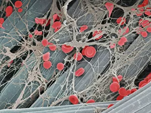
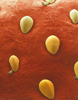
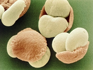
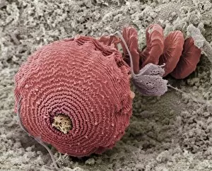
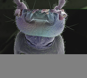
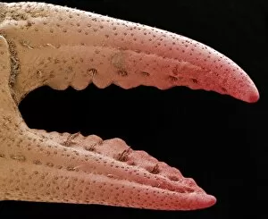
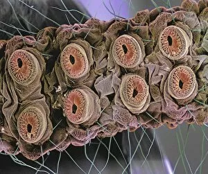
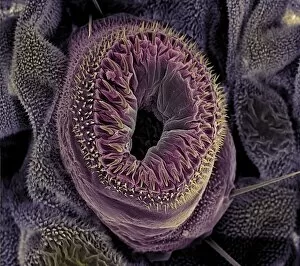
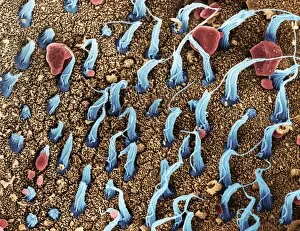
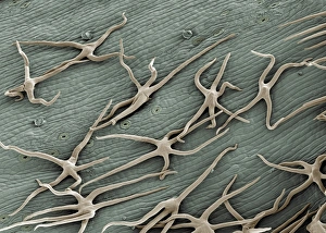
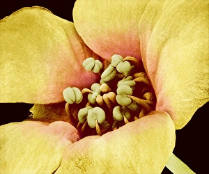
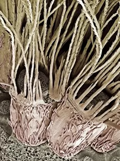
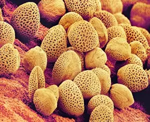
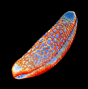
"Exploring the Intricate World of Coloured SEM: From Blood Clots to Fly Eyes" Delving into the fascinating realm of coloured scanning electron microscopy (SEM), we uncover a myriad of captivating images that offer a unique perspective on various subjects. In one striking image, we observe a blood clot delicately resting on a plaster surface, revealing intricate details that would otherwise go unnoticed. The SEM technique allows us to zoom in and examine this microscopic phenomenon with awe-inspiring clarity. Moving on to nature's wonders, we encounter the velvety surface of a strawberry fruit under SEM. Each tiny bump and groove becomes magnified, showcasing the extraordinary beauty hidden within even the simplest objects. Venturing further into botanical marvels, pollen grains from a Scots Pine tree come alive through vivid colours and intricate patterns when viewed under SEM. These minuscule structures take center stage as they reveal their role in plant reproduction. Shifting gears towards materials science, high-density foam manufactured by Zotefoam takes on an otherworldly appearance when observed using coloured SEM. Its cellular structure is brought to life with vibrant hues and reveals its potential applications in insulation and packaging industries. Zooming back into the world of living organisms, we encounter mesmerizing images of protozoans captured through SEM. These single-celled creatures showcase their complex forms and highlight their significance in ecological systems. Continuing our exploration underwater, we delve into the world of shore crabs through coloured SEM imagery. Their exoskeletons become works of art as every ridge and spine is meticulously showcased for our admiration. The head of an earwig becomes an enigmatic masterpiece when examined at close range using coloured SEM techniques. Its intricate features are unveiled with astonishing precision - an ode to nature's remarkable design skills. A crab pincer takes center stage as it undergoes scrutiny under coloured SEM; its serrated edges appear like finely crafted weapons designed for survival in harsh marine environments.