Canvas Print > Fine Art Storehouse > Photo Libraries > Dorling Kindersley Prints
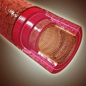
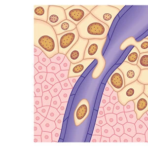
Canvas Print : Cross section illustration of human small intestine showing muscle layer, villus and plicae


![]()
Canvas Prints from Fine Art Storehouse
Cross section illustration of human small intestine showing muscle layer, villus and plicae
Unleash your creativity and transform your space into a visual masterpiece!
Dorling Kindersley
Media ID 13555833
© This content is subject to copyright
Anatomy Biomedical Illustration Blood Flow Connection Cross Section Human Digestive System Human Intestine Human Muscle Human Vein Layer Physiology Small Tiered Human Body Part Mesentery Serosa Villus Watercolour Painting
30"x20" (76x51cm) Canvas Print
Discover the intricacies of the human body with our stunning Canvas Print of a cross section illustration of the small intestine by Dorling Kindersley from the Fine Art Storehouse collection. This captivating work of art showcases the intricate details of the muscle layer, villus, and plicae in the small intestine, bringing a unique blend of science and art to your home or office. The high-quality canvas print offers vibrant colors and impressive detail, ensuring a beautiful addition to any space. Explore the depths of the human anatomy and ignite your curiosity with this exquisite piece from Media Storehouse.
Delivered stretched and ready to hang our premium quality canvas prints are made from a polyester/cotton blend canvas and stretched over a 1.25" (32mm) kiln dried knot free wood stretcher bar. Packaged in a plastic bag and secured to a cardboard insert for safe transit.
Canvas Prints add colour, depth and texture to any space. Professionally Stretched Canvas over a hidden Wooden Box Frame and Ready to Hang
Estimated Product Size is 76.2cm x 50.8cm (30" x 20")
These are individually made so all sizes are approximate
Artwork printed orientated as per the preview above, with landscape (horizontal) orientation to match the source image.
EDITORS COMMENTS
This watercolor print by Dorling Kindersley offers a detailed cross-section illustration of the human small intestine. The artwork showcases the intricate layers and structures that make up this vital organ within our digestive system. From the outer serosa to the inner submucosa, every layer is meticulously depicted, highlighting their unique functions. The artist's skillful use of watercolors brings life to this scientific illustration, with vibrant hues contrasting against a crisp white background. The high-angle view allows us to appreciate the tiered arrangement of plicae circulares, which enhance nutrient absorption in the intestines. Each individual villus is delicately painted, emphasizing their role in increasing surface area for efficient digestion. The presence of veins throughout the muscle layer reminds us of the essential blood flow required for proper functioning. Mesentery, connecting and supporting these structures, can also be observed in this stunning piece. Without any people present, this print solely focuses on capturing the beauty and complexity of our internal anatomy. It serves as an educational tool for students studying physiology or biomedical sciences while also appealing to those fascinated by anatomical art. Overall, Dorling Kindersley's masterful depiction provides both aesthetic pleasure and valuable knowledge about our remarkable human body.
MADE IN AUSTRALIA
Safe Shipping with 30 Day Money Back Guarantee
FREE PERSONALISATION*
We are proud to offer a range of customisation features including Personalised Captions, Color Filters and Picture Zoom Tools
SECURE PAYMENTS
We happily accept a wide range of payment options so you can pay for the things you need in the way that is most convenient for you
* Options may vary by product and licensing agreement. Zoomed Pictures can be adjusted in the Cart.