Jigsaw Puzzle > Fine Art Storehouse > Photo Libraries > Dorling Kindersley Prints
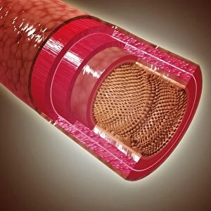
Jigsaw Puzzle : Cross section illustration of human small intestine showing muscle layer, villus and plicae


![]()
Jigsaw Puzzles from Fine Art Storehouse
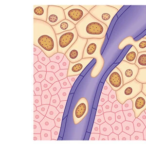
Cross section illustration of human small intestine showing muscle layer, villus and plicae
Unleash your creativity and transform your space into a visual masterpiece!
Dorling Kindersley
Media ID 13555833
© This content is subject to copyright
Anatomy Biomedical Illustration Blood Flow Connection Cross Section Human Digestive System Human Intestine Human Muscle Human Vein Layer Physiology Small Tiered Human Body Part Mesentery Serosa Villus Watercolour Painting
Jigsaw Puzzle (1000 Pieces)
Discover the wonders of the human body with our educational Jigsaw Puzzle from Media Storehouse and Dorling Kindersley's Fine Art Storehouse. This intricately detailed puzzle depicts a cross-section illustration of the small intestine, allowing you to explore the complex structure of this essential organ. Delve deeper into the anatomy of the muscle layer, villus, and plicae as you piece together this captivating puzzle. A perfect addition to any home or classroom, this puzzle is not only fun but also an excellent way to learn and expand your knowledge of the human body.
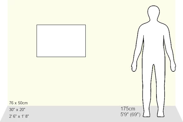
1000 piece puzzles are custom made in Australia and hand-finished on 100% recycled 1.6mm thick laminated puzzle boards. There is a level of repetition in jigsaw shapes with each matching piece away from its pair. The completed puzzle measures 76x50cm and is delivered packaged in an attractive presentation box specially designed to fit most mail slots with a unique magnetic lid
Jigsaw Puzzles are an ideal gift for any occasion
Estimated Product Size is 76cm x 50.2cm (29.9" x 19.8")
These are individually made so all sizes are approximate
Artwork printed orientated as per the preview above, with landscape (horizontal) or portrait (vertical) orientation to match the source image.
EDITORS COMMENTS
This watercolor print by Dorling Kindersley offers a detailed cross-section illustration of the human small intestine. The artwork showcases the intricate layers and structures that make up this vital organ within our digestive system. From the outer serosa to the inner submucosa, every layer is meticulously depicted, highlighting their unique functions. The artist's skillful use of watercolors brings life to this scientific illustration, with vibrant hues contrasting against a crisp white background. The high-angle view allows us to appreciate the tiered arrangement of plicae circulares, which enhance nutrient absorption in the intestines. Each individual villus is delicately painted, emphasizing their role in increasing surface area for efficient digestion. The presence of veins throughout the muscle layer reminds us of the essential blood flow required for proper functioning. Mesentery, connecting and supporting these structures, can also be observed in this stunning piece. Without any people present, this print solely focuses on capturing the beauty and complexity of our internal anatomy. It serves as an educational tool for students studying physiology or biomedical sciences while also appealing to those fascinated by anatomical art. Overall, Dorling Kindersley's masterful depiction provides both aesthetic pleasure and valuable knowledge about our remarkable human body.
MADE IN AUSTRALIA
Safe Shipping with 30 Day Money Back Guarantee
FREE PERSONALISATION*
We are proud to offer a range of customisation features including Personalised Captions, Color Filters and Picture Zoom Tools
SECURE PAYMENTS
We happily accept a wide range of payment options so you can pay for the things you need in the way that is most convenient for you
* Options may vary by product and licensing agreement. Zoomed Pictures can be adjusted in the Cart.