
Canvas Print : Babys nail, SEM


![]()
Canvas Prints from Science Photo Library
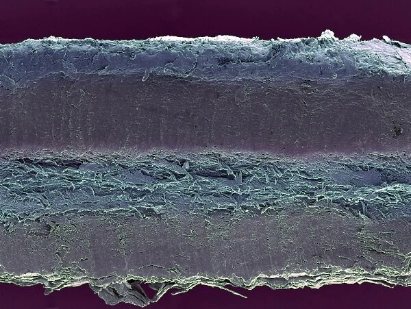
Babys nail, SEM
Babys nail. Coloured scanning electron micrograph (SEM) of a section through a babys nail. Nails are keratinised structures (they contain the protein keratin) made up of dead tissue bound together to form a hard, protective structure on the tips of the fingers and toes. The growth of a fingernail is similar to that of a hair, another keratinised structure that grows on a human body. Nails form a layered structure, with the bottom layer attached to the skin. Several layers are seen here
Science Photo Library features Science and Medical images including photos and illustrations
Media ID 6452445
© STEVE GSCHMEISSNER/SCIENCE PHOTO LIBRARY
Baby Development False Colour Finger Nail Infant Keratin Layer Layers Nail Young False Coloured Section Sectioned
20"x16" (51x41cm) Canvas Print
Discover the intricacies of life with Media Storehouse's Canvas Prints featuring "Babys nail, SEM" by Science Photo Library. This captivating image offers a unique perspective into the world of science, showcasing a coloured scanning electron micrograph (SEM) of a section through a baby's nail. Delve into the complexities of keratinised structures and add a touch of scientific wonder to your home or office decor. Our high-quality canvas prints are meticulously crafted to bring out the vibrant colours and intricate details of this mesmerising SEM image, ensuring a stunning addition to any space.
Delivered stretched and ready to hang our premium quality canvas prints are made from a polyester/cotton blend canvas and stretched over a 1.25" (32mm) kiln dried knot free wood stretcher bar. Packaged in a plastic bag and secured to a cardboard insert for safe transit.
Canvas Prints add colour, depth and texture to any space. Professionally Stretched Canvas over a hidden Wooden Box Frame and Ready to Hang
Estimated Product Size is 50.8cm x 40.6cm (20" x 16")
These are individually made so all sizes are approximate
Artwork printed orientated as per the preview above, with landscape (horizontal) orientation to match the source image.
EDITORS COMMENTS
This print showcases the intricate details of a baby's nail, captured through a scanning electron microscope (SEM). The image reveals the colorful layers that make up this keratinized structure, which serves as a protective barrier on the fingertips and toes. Just like hair, nails are composed of dead tissue bound together by the protein keratin. The photograph provides an insightful glimpse into the development and anatomy of human nails during early stages of life. It highlights how these keratinous structures grow in layers, with the bottom layer firmly attached to the skin. The SEM technique allows us to appreciate the fine textures and biological complexity present in even seemingly ordinary parts of our bodies. Through this image, we gain a deeper understanding of normal nail formation and healthy infant development. It reminds us that every aspect of our anatomy is intricately designed for specific functions. This scientific marvel not only captivates biologists but also sparks curiosity among those interested in exploring our own biology at microscopic levels. Science Photo Library has once again provided us with an awe-inspiring visual representation that merges artistry with scientific exploration. This stunning photograph invites viewers to appreciate both its aesthetic beauty and its potential for advancing our knowledge about human anatomy.
MADE IN AUSTRALIA
Safe Shipping with 30 Day Money Back Guarantee
FREE PERSONALISATION*
We are proud to offer a range of customisation features including Personalised Captions, Color Filters and Picture Zoom Tools
SECURE PAYMENTS
We happily accept a wide range of payment options so you can pay for the things you need in the way that is most convenient for you
* Options may vary by product and licensing agreement. Zoomed Pictures can be adjusted in the Cart.