
Photoreceptors Collection
Photoreceptors, specifically rod and cone cells of the eye, play a crucial role in our visual perception


All Professionally Made to Order for Quick Shipping
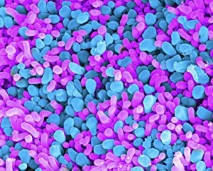
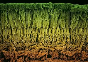
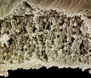
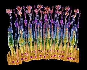
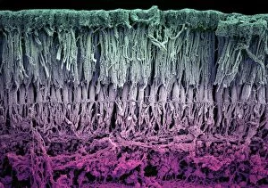
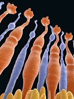
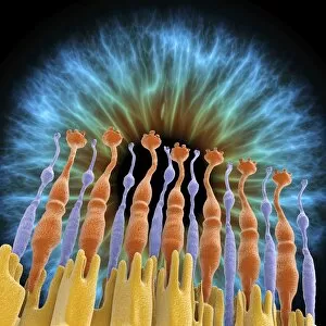
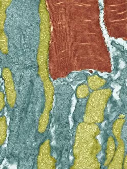
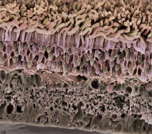
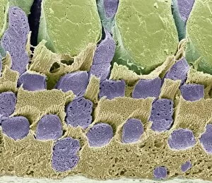
Photoreceptors, specifically rod and cone cells of the eye, play a crucial role in our visual perception. These microscopic structures are responsible for converting light into electrical signals that can be interpreted by the brain. In SEM images such as C014 / 4866 and C014 / 4864, we get a close-up view of these photoreceptor cells. The intricate details captured in these pictures showcase their unique shapes and structures. Rod cells are elongated with a cylindrical shape, while cone cells have a more conical appearance. Artwork like C017 / 7791 beautifully illustrates the arrangement of rods and cones on the retina. This artwork provides an artistic representation of how these they can distributed across this delicate tissue at the back of our eyes. Pictures No. 11675579, 10876996, 10876993, 10876995, and 10876994 further emphasize the importance in vision. They give us glimpses into their location within the retina and highlight their distinct characteristics. The images F008 /0713, F008/0719, F008/0714, and F008/0712 provide additional insights into the structure of retinal tissue containing rod and cone cells. These high-resolution photographs offer detailed views that help researchers better understand how these photoreceptors function together to enable sight. Overall, studying photoreceptors is essential for unraveling the mysteries behind human vision. Through advanced imaging techniques like scanning electron microscopy (SEM) or detailed artworks capturing their arrangement on the retina's surface; scientists continue to explore these remarkable cellular components that allow us to perceive color, depth, motion - ultimately shaping our visual experiences every day.