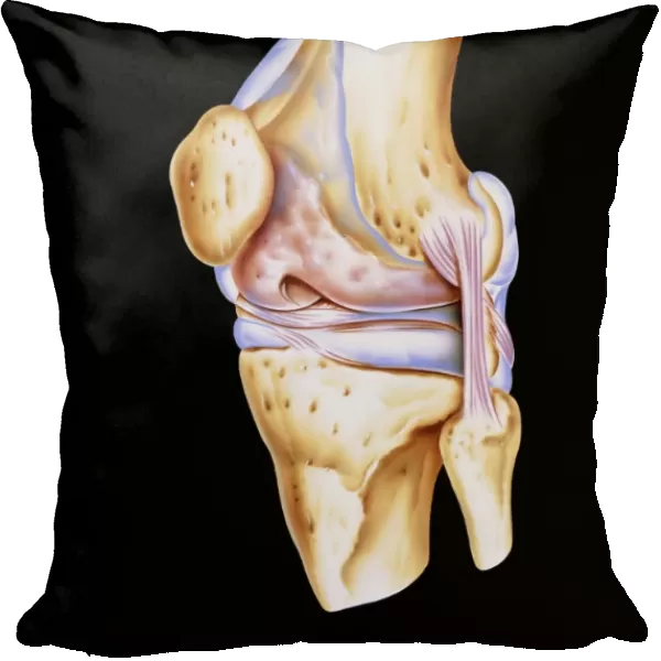
Cushion : Artwork of bones & ligaments in human knee joint
![]()

Home Decor from Science Photo Library
Artwork of bones & ligaments in human knee joint
Knee joint. Illustration showing bones and tendons of the human knee (genual) joint. The knee is the largest joint in the human body. It forms the point of articulation between the upper leg bone (femur, at top) and the two bones of the lower limb (tibia at lower left, fibula at lower right). The articulation surface is formed from condyles (round bone ends) between the femur and tibia. These condyles are covered in cartilage (white) to prevent friction on movement. Ligaments (pink) fix this joint together: a fibular collateral ligament attaches the fibula to femur; cruciate ligaments attach tibia to femur between the condyles. The oval knee cap bone (patella) protects the joint
Science Photo Library features Science and Medical images including photos and illustrations
Media ID 6419922
© JOHN BAVOSI/SCIENCE PHOTO LIBRARY
Bones Joint Knee Knee Joint Ligament Patella
Cushion
Refresh your home decor with a beautiful full photo 16"x16" (40x40cm) cushion, complete with cushion pad insert. Printed on both sides and made from 100% polyester with a zipper on the bottom back edge of the cushion cover. Care Instructions: Warm machine wash, do not bleach, do not tumble dry. Warm iron inside out. Do not dry clean.
Accessorise your space with decorative, soft cushions
Estimated Product Size is 40cm x 40cm (15.7" x 15.7")
These are individually made so all sizes are approximate
Artwork printed orientated as per the preview above, with landscape (horizontal) or portrait (vertical) orientation to match the source image.
EDITORS COMMENTS
This print showcases the intricate artwork of bones and ligaments in a human knee joint. The knee joint, known as the genual joint, is depicted with utmost precision and detail. As the largest joint in the human body, it serves as the crucial point of articulation between the upper leg bone (femur) and the two lower limb bones (tibia and fibula). The artistry beautifully captures the condyles, which are rounded ends of bones found between the femur and tibia. These condyles play a vital role in facilitating movement while being protected by a layer of white cartilage that prevents friction. In this masterpiece, pink ligaments can be seen firmly holding this complex joint together. A fibular collateral ligament connects the fibula to femur while cruciate ligaments attach both tibia and femur between these condyles. Adding to its elegance is an oval-shaped bone called patella or kneecap that acts as a protective shield for this remarkable joint. Science Photo Library has skillfully merged science with art through this print, allowing us to appreciate not only our anatomy but also its aesthetic beauty. It serves as a reminder of how intricately designed our bodies are, showcasing both strength and vulnerability within one frame.
MADE IN AUSTRALIA
Safe Shipping with 30 Day Money Back Guarantee
FREE PERSONALISATION*
We are proud to offer a range of customisation features including Personalised Captions, Color Filters and Picture Zoom Tools
SECURE PAYMENTS
We happily accept a wide range of payment options so you can pay for the things you need in the way that is most convenient for you
* Options may vary by product and licensing agreement. Zoomed Pictures can be adjusted in the Cart.


