Cushion : False-colour SEM of dorsal surface of tongue


![]()
Home Decor from Science Photo Library
False-colour SEM of dorsal surface of tongue
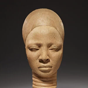
Tongue surface. False-colour scanning electron micrograph (SEM) of the dorsal surface of the tongue. It is covered by two types of projections known as filiform and fungiform papillae. Filiform papillae, also known as conical papillae, are more numerous and have mechanical and tactile functions. They form a rough surface which helps the mastication process. Fungiform papillae are round and bigger than filiform papillae. On many fungiform papillae taste buds are found. Magnification: x25 at 6x7cm size. Magnification: x35 at 4x5 inch size
Science Photo Library features Science and Medical images including photos and illustrations
Media ID 6422526
© PROF. P. MOTTA/DEPT. OF ANATOMY/UNIVERSITY LA SAPIENZA , ROME/SCIENCE PHOTO LIBRARY
Filiform Papilla Fungiform Papilla Magnified Image Microscopic Photos Papilla Papillae Subjects Tongue False Coloured
Cushion
Refresh your home decor with a beautiful full photo 16"x16" (40x40cm) cushion, complete with cushion pad insert. Printed on both sides and made from 100% polyester with a zipper on the bottom back edge of the cushion cover. Care Instructions: Warm machine wash, do not bleach, do not tumble dry. Warm iron inside out. Do not dry clean.
Accessorise your space with decorative, soft cushions
Estimated Product Size is 40cm x 40cm (15.7" x 15.7")
These are individually made so all sizes are approximate
Artwork printed orientated as per the preview above, with landscape (horizontal) or portrait (vertical) orientation to match the source image.
EDITORS COMMENTS
This print showcases the intricate details of the dorsal surface of a tongue, captured using a false-colour scanning electron microscope (SEM). The image reveals two distinct types of projections known as filiform and fungiform papillae. The filiform papillae, also referred to as conical papillae, are abundant in number and serve mechanical and tactile functions. They create a rough surface that aids in the process of mastication or chewing. On the other hand, the larger round structures seen in this image are fungiform papillae. These specialized projections house taste buds on many of their surfaces. With a magnification level of x25 at 6x7cm size and x35 at 4x5 inch size, this microscopic photograph offers an up-close look into the fascinating world within our mouths. It provides valuable insights into the anatomy and composition of human tongues. This stunning visual representation not only highlights scientific subjects such as tongue anatomy but also showcases how technology can unveil hidden wonders within our bodies. This particular print is part of Science Photo Library's extensive collection featuring various microscopic photos capturing different aspects of life sciences.
MADE IN AUSTRALIA
Safe Shipping with 30 Day Money Back Guarantee
FREE PERSONALISATION*
We are proud to offer a range of customisation features including Personalised Captions, Color Filters and Picture Zoom Tools
SECURE PAYMENTS
We happily accept a wide range of payment options so you can pay for the things you need in the way that is most convenient for you
* Options may vary by product and licensing agreement. Zoomed Pictures can be adjusted in the Cart.