Home > Arts > Artists > S > George Stubbs
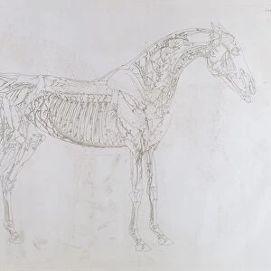
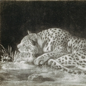
Study of a Tiger, Lateral View, Abdominal Viscera Exposed, from A Comparative Anatomical Exposition of the Structure of the Human Body with that of a Tiger and a Common Fowl, c.1795-1806 (graphite on cartridge paper)
")
![]()
Wall Art and Photo Gifts from Fine Art Finder
Study of a Tiger, Lateral View, Abdominal Viscera Exposed, from A Comparative Anatomical Exposition of the Structure of the Human Body with that of a Tiger and a Common Fowl, c.1795-1806 (graphite on cartridge paper)
XYC261309 Study of a Tiger, Lateral View, Abdominal Viscera Exposed, from A Comparative Anatomical Exposition of the Structure of the Human Body with that of a Tiger and a Common Fowl, c.1795-1806 (graphite on cartridge paper) by Stubbs, George (1724-1806); 40.6x53.7 cm; Yale Center for British Art, Paul Mellon Collection, USA
Media ID 33069110
© Bridgeman Images
Anatomical Anatomist Biological Biology Diagram Domestic Cat George Stubbs Internal Anatomy Kitten Kittens Legs Limbs Proportion Recto Series Skeletal Skeleton Skull Stubbs George 1724 1806 Veterinary Britisch Britisch Egnland
FEATURES IN THESE COLLECTIONS
> Animals
> Mammals
> Cats (Domestic)
> Related Images
> Animals
> Mammals
> Cats (Wild)
> Tiger
> Animals
> Wild
> Big Cats
> Tiger
> Arts
> Artists
> S
> George Stubbs
> Arts
> Realistic drawings
> Graphite art
> Fine art
> Europe
> United Kingdom
> Northern Ireland
> Posters
> Europe
> United Kingdom
> Northern Ireland
> Related Images
> Fine Art Finder
> Artists
> George Stubbs
> Sport
> Sports Stars
> Paul George
EDITORS COMMENTS
This print captures a detailed study of a tiger, showcasing its lateral view with the abdominal viscera exposed. Created by George Stubbs between 1795 and 1806, this graphite on cartridge paper artwork is part of his larger project titled "A Comparative Anatomical Exposition of the Structure of the Human Body with that of a Tiger and a Common Fowl". Measuring at 40.6x53.7 cm, this piece can be found in the prestigious Yale Center for British Art's Paul Mellon Collection in the United States. The artwork belongs to the 18th century, also known as the xviii or c18th century. Stubbs was not only an accomplished artist but also an anatomist who dedicated himself to exploring internal anatomy through his art. This particular work showcases his meticulous attention to detail and scientific accuracy. The image depicts a domestic cat-like animal in all its glory, revealing its intricate biology and internal structures. It serves as both an artistic masterpiece and a valuable resource for medical professionals studying feline anatomy. George Stubbs' contribution to both art and science cannot be overstated. His works continue to inspire awe even centuries after their creation, bridging the gap between artistic expression and scientific exploration. Richard Caspole has skillfully captured this remarkable piece through photography, allowing viewers around the world to appreciate Stubbs' genius from Fine Art Finder's collection without commercial use intentions.
MADE IN AUSTRALIA
Safe Shipping with 30 Day Money Back Guarantee
FREE PERSONALISATION*
We are proud to offer a range of customisation features including Personalised Captions, Color Filters and Picture Zoom Tools
SECURE PAYMENTS
We happily accept a wide range of payment options so you can pay for the things you need in the way that is most convenient for you
* Options may vary by product and licensing agreement. Zoomed Pictures can be adjusted in the Cart.