Framed Print > Science > SEM
Framed Print : Coloured SEM of a fungiform papilla of the tongue


![]()
Framed Photos from Science Photo Library
Coloured SEM of a fungiform papilla of the tongue
Fungiform papilla of tongue. Coloured scanning electron micrograph (SEM) of a fungiform papilla of the tongue. The taste buds, the organs of taste, are found under the fungiform papillae. Fungiform papillae are covered by layers of stratified squamous epithelium, which constantly shed their dead cells (seen here as thin flakes) and replace them with cells from their underlying layers. Magnification unknown
Science Photo Library features Science and Medical images including photos and illustrations
Media ID 6422550
© STEVE GSCHMEISSNER/SCIENCE PHOTO LIBRARY
Epithelium Fungiform Fungiform Papilla Papilla Squamous Squamous Epithelium Tongue
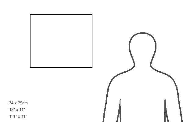
13.5"x11.5" (34x29cm) Premium Frame
Discover the intricacies of the human body with our captivating Framed Prints from Media Storehouse. This striking Coloured SEM of a fungiform papilla of the tongue, sourced from Science Photo Library, offers a mesmerizing glimpse into the microscopic world of taste. Each print showcases the vibrant details of the fungiform papilla, home to taste buds that bring flavors to life. Elevate your home or office décor with this scientific masterpiece, sure to spark curiosity and conversation.
Framed and mounted 9x7 print. Professionally handmade full timber moulded frames are finished off with framers tape and come with a hanging solution on the back. Outer dimensions are 13.5x11.5 inches (34x29cm). Quality timber frame frame moulding (20mm wide and 30mm deep) with frame colours in your choice of black, white, or raw oak and a choice of black or white card mounts. Frames have a perspex front providing a virtually unbreakable glass-like finish which is easily cleaned with a damp cloth.
Contemporary Framed and Mounted Prints - Professionally Made and Ready to Hang
Estimated Image Size (if not cropped) is 21.4cm x 21.4cm (8.4" x 8.4")
Estimated Product Size is 34cm x 29.2cm (13.4" x 11.5")
These are individually made so all sizes are approximate
Artwork printed orientated as per the preview above, with landscape (horizontal) or portrait (vertical) orientation to match the source image.
EDITORS COMMENTS
This print showcases the intricate details of a fungiform papilla, one of the many fascinating structures on our tongue. Through the lens of a scanning electron microscope (SEM), this coloured image reveals the complexity and beauty hidden within our taste buds. The fungiform papillae play a crucial role in our sense of taste, housing the organs responsible for detecting different flavors. Beneath their surface lies an array of taste buds, which are essential for experiencing sweet, sour, salty, bitter, and umami sensations. Covered by layers of stratified squamous epithelium, these papillae constantly undergo renewal as dead cells flake off and are replaced by fresh ones from below. This perpetual process ensures that our taste receptors remain functional and responsive to new culinary experiences. While we don't know the exact magnification used to capture this stunning image, it's clear that it offers us a glimpse into the microscopic wonders within our own bodies. The intricacy displayed here reminds us just how remarkable human anatomy truly is. This print from Science Photo Library serves as a testament to both scientific curiosity and artistic appreciation for nature's marvels. It invites us to explore further into the realms unseen with awe and wonderment at every turn.
MADE IN AUSTRALIA
Safe Shipping with 30 Day Money Back Guarantee
FREE PERSONALISATION*
We are proud to offer a range of customisation features including Personalised Captions, Color Filters and Picture Zoom Tools
SECURE PAYMENTS
We happily accept a wide range of payment options so you can pay for the things you need in the way that is most convenient for you
* Options may vary by product and licensing agreement. Zoomed Pictures can be adjusted in the Cart.