Fraud Blocker Skip to main content

Actin Collection

EDITORS COMMENTS"Actin: The Dynamic Force Behind Cell Structure and Muscle Contraction" From HeLa cells to muscle fibers

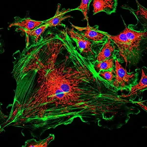
Background imageActin Collection: HeLa cells, light micrograph C017 / 8299

HeLa cells, light micrograph C017 / 8299
HeLa cells, multiphoton fluorescence micrograph (MFM). The cell nuclei, which contain the cells genetic information, are purple. Microtubules are blue and actin microfilaments are red

Background imageActin Collection: Cell structure

Cell structure. Confocal light micrograph of cultured endothelial cells. A fluorescent dye has been used to show the cell structure

Background imageActin Collection: HeLa cells, light micrograph C013 / 4774

HeLa cells, light micrograph C013 / 4774
HeLa cells. Multi-photon fluorescence light micrograph of a group of cultured HeLa cells, showing the cell nuclei, which contain the cells genetic information (DNA, red)

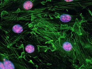
Background imageActin Collection: Immunofluorescent LM of fibroblast cell nuclei

Immunofluorescent LM of fibroblast cell nuclei
Cell nuclei. Immunofluorescent light micrograph of cultured fibroblast cells showing nuclei with " nucleolar necklaces"

Background imageActin Collection: Intestinal microvilli, TEM

Intestinal microvilli, TEM
Intestinal microvilli, coloured transmission electron micrograph (TEM)

Background imageActin Collection: Heart muscle, confocal light micrograph

Heart muscle, confocal light micrograph
Heart muscle. Confocal light micrograph of a section through cardiac (heart) muscle

Structure Skeletal Muscle. myofibril with thin and thick filament. close up of a sarcomere. Muscles contract by sliding the myosin and actin filaments Structure Skeletal Muscle

Background imageActin Collection: Each skeletal muscle fiber has many bundles of myofilaments

Each skeletal muscle fiber has many bundles of myofilaments. Each bundle is called a myofibril

Background imageActin Collection: Muscle fibre structure, artwork

Muscle fibre structure, artwork
Muscle fibre. Computer artwork showing the structure of a muscle cell, also known as a muscle fibre. The cell is surrounded by a plasma membrane called the sarcolemma (cream)

Background imageActin Collection: Smooth muscle tissue, TEM

Smooth muscle tissue, TEM
Smooth muscle tissue. Transmission electron micrograph (TEM) of a longitudinal section through smooth muscle cells

Background imageActin Collection: Illustration of muscle contraction

Illustration of muscle contraction

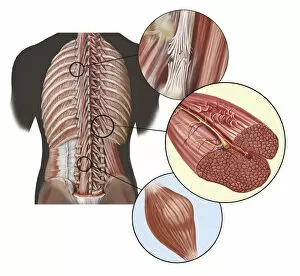
Background imageActin Collection: Detail of deep back muscles with a close-up of sprain, strain and spasm

Detail of deep back muscles with a close-up of sprain, strain and spasm

Background imageActin Collection: Illustration of muscle contraction (with labels)

Illustration of muscle contraction (with labels)

Background imageActin Collection: Neurofibromatosis, a genetic disorder of the nervous system

Neurofibromatosis, a genetic disorder of the nervous system
A genetic disorder of the nervous system, neurofibromatosis causes tumors to form on nerves throughout the body

Background imageActin Collection: Torn muscle fibers with healing stages surrounding

Torn muscle fibers with healing stages surrounding. 1. Macrophage clean-up. 2. New cells migrate to repair. 3. Cells differentiate to become muscle cells

Background imageActin Collection: Embryonic smooth muscle cell C018 / 8595

Embryonic smooth muscle cell C018 / 8595
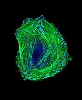
Embryonic smooth muscle cell, immunofluorescence micrograph. Actin filaments are green, the protein vinculin is light blue. Actin is part of the cytoskeleton

Background imageActin Collection: Beta-actin molecule F006 / 9358

Beta-actin molecule F006 / 9358
Beta-actin. Molecular model of the cytoskeletal protein beta-actin bound to profilin

Background imageActin Collection: DNase I complex, molecular model F006 / 9239

DNase I complex, molecular model F006 / 9239
DNase I complex. Molecular model of deoxyribonuclease I complexed with actin

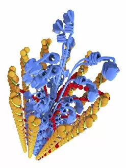
Background imageActin Collection: Actin Myosin Muscle Model, artwork C014 / 2661

Actin Myosin Muscle Model, artwork C014 / 2661
Computer artwork of the molecular actin myosin muscle structure

Background imageActin Collection: Muscle contraction proteins

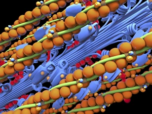
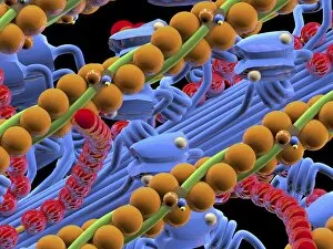
Muscle contraction proteins. Molecular model of muscle protein motor cross-bridges during contraction in muscle. The cross-bridge is seen from the side, with contraction taking place horizontally

Background imageActin Collection: Neuromuscular junction, artwork

Neuromuscular junction, artwork
Neuromuscular junction. Computer artwork showing the juntion between a neuron (nerve cell, light blue) and a muscle cell, known as a neuromuscular junction

Background imageActin Collection: Actin Myosin Muscle Model, artwork C014 / 2658

Actin Myosin Muscle Model, artwork C014 / 2658
Computer artwork of the molecular actin myosin muscle structure

Background imageActin Collection: Actin Myosin Muscle Model, artwork C014 / 2659

Actin Myosin Muscle Model, artwork C014 / 2659
Computer artwork of the molecular actin myosin muscle structure

Background imageActin Collection: Actin myosin muscle model, artwork C014 / 2660

Actin myosin muscle model, artwork C014 / 2660
Computer artwork of the molecular actin myosin muscle structure

Background imageActin Collection: Human muscle fibres, diagram

Human muscle fibres, diagram
Human muscle fibres. Diagram showing the structure of human muscles from the cellular to the macroscopic level

Background imageActin Collection: Kidney tissue, fluorescence micrograph C016 / 8484

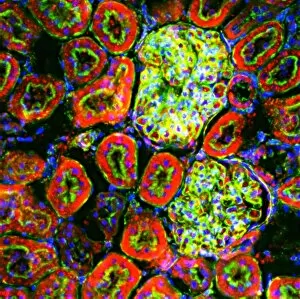
Kidney tissue, fluorescence micrograph C016 / 8484
Kidney tissue. Fluorescence deconvolution micrograph of a section through a kidney, showing glomeruli (green), cell nuclei (blue dots), and renal tubules (red, circular)

Background imageActin Collection: Heart muscle, fluorescence micrograph C016 / 8483

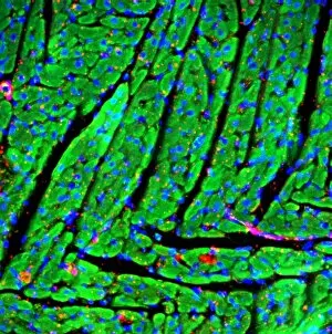
Heart muscle, fluorescence micrograph C016 / 8483
Heart muscle. Fluorescence deconvolution micrograph of a section through heart tissue, showing the angular distribution of the myocardium (cardiac muscle) fibres (green)

Background imageActin Collection: Heart muscle, fluorescence micrograph C016 / 8481

Heart muscle, fluorescence micrograph C016 / 8481
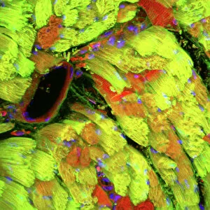
Heart muscle. Fluorescence deconvolution micrograph of a section through heart tissue, showing blood vessels (oval, centre-left and upper centre) running through the myocardium (cardiac muscle)

Background imageActin Collection: Heart muscle, fluorescence micrograph C016 / 8478

Heart muscle, fluorescence micrograph C016 / 8478
Heart muscle. Fluorescence deconvolution micrograph of a section through heart tissue, showing a blood vessel (diagonal, centre) running through the myocardium (cardiac muscle, green)

Background imageActin Collection: Heart muscle, fluorescence micrograph C016 / 8477

Heart muscle, fluorescence micrograph C016 / 8477
Heart muscle. Fluorescence deconvolution micrograph of a section through heart tissue, showing a blood vessel (blue, diagonal, centre) running through the myocardium (cardiac muscle, green)

Background imageActin Collection: Adrenal gland, fluorescence micrograph C016 / 8472

Adrenal gland, fluorescence micrograph C016 / 8472
Adrenal gland. Fluorescence deconvolution micrograph of a section through an adrenal gland, showing the zona glomerulosa and zona fasciculata

Background imageActin Collection: Fibroblast cell, fluorescent micrograph

Fibroblast cell, fluorescent micrograph
Fibroblast cell. Confocal laser scanning micrograph of a fibroblast cell. The cell nucleus, which contains the cells genetic information, is green

Background imageActin Collection: HeLa cells, light micrograph C013 / 4773

HeLa cells, light micrograph C013 / 4773
HeLa cells. Multi-photon fluorescence light micrograph of a group of cultured HeLa cells, showing the cell nuclei, which contain the cells genetic information (DNA, blue), and microtubules (pink)

Background imageActin Collection: Dividing cell, light micrograph

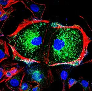
Dividing cell, light micrograph
Cell division. Fluorescent light micrograph of a cell that has divided by mitosis, the asexual replication of a cell into two new cells

Background imageActin Collection: Fibroblast cells, light micrograph

Fibroblast cells, light micrograph
Fibroblast cells. Confocal light micrograph of human fibroblast cells. The cell nuclei, which contain the cells genetic information, are red

Background imageActin Collection: Rat tongue, light micrograph

Rat tongue, light micrograph
Rat tongue. Confocal light micrograph of an en face section through a rats tongue, showing tastebuds. Actin protein filaments, which make up part of the cytoskeleton, are orange

Background imageActin Collection: Retina blood vessel and nerve cells

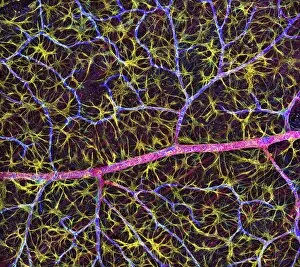
Retina blood vessel and nerve cells
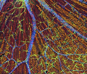
Retina cells. Fluorescent light micrograph of cells in the retina, the light-sensitive membrane that lines the back of the eyeball

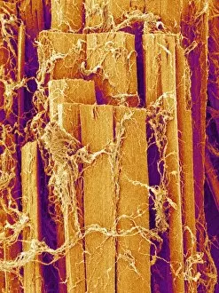
Background imageActin Collection: Skeletal muscle fibre

Skeletal muscle fibre. Coloured scanning electron micrograph (SEM) of skeletal muscle fibre. This type of muscle is striated

Background imageActin Collection: Skeletal muscle, TEM

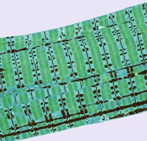
Skeletal muscle, TEM
Skeletal muscle. Coloured transmission electron micrograph (TEM) of a longitudinal section through skeletal, or striated, muscle

Background imageActin Collection: Nerve cell growth

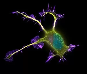
Nerve cell growth. Fluorescent light micrograph of a PC12 cell following stimulation by nerve growth factor. The cell body contains the nucleus (green)

Background imageActin Collection: Muscle fibres

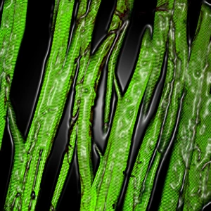
Muscle fibres. Computer-enhanced confocal light micrograph of skeletal (striated) muscle fibres. Skeletal muscle is under the conscious control of the brain

Background imageActin Collection: Artwork showing structure of human skeletal muscle

Artwork showing structure of human skeletal muscle
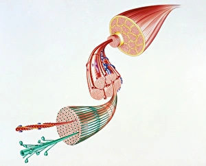
Skeletal muscle. Illustration of the structure of human skeletal muscle. Skeletal muscle (top right) consists of bundles of long, thin cells called muscle fibres

Background imageActin Collection: Cultured cell, light micrograph

Cultured cell, light micrograph
Cultured cell. Immunofluorescent light micrograph of an epithelial cell (round, upper right). The cell proteins are marked by fluorescent dyes

Background imageActin Collection: Human epithelial cells

Human epithelial cells. Fluorescent light micrograph of human epithelial cells in culture. The nuclei, which contain the cells genetic information DNA (deoxyribonucleic acid), have been dyed blue

Background imageActin Collection: Muscle cell anatomy, artwork

Muscle cell anatomy, artwork
Muscle cell anatomy. Artwork of the anatomy of a muscle cell, also known as a muscle fibre. The cell is surrounded by a plasma membrane called the sarcolemma (grey)

Background imageActin Collection: Muscle structure, artwork

Muscle structure, artwork
Muscle structure. Computer artwork showing the protein structure of myofibrils and how they are bound together to form muscle fibres

Background imageActin Collection: Cytoskeleton components, artwork

Cytoskeleton components, artwork. Three main components of the cytoskeleton, the internal support structure of a cell, are shown here

Background imageActin Collection: Cytoskeleton components, diagram

Cytoskeleton components, diagram. The cytoskeleton is the internal support structure of a cell, composed of filaments of various diameters in nanometres (nm)

Background imageActin Collection: Kidney cells, light micrograph

Kidney cells, light micrograph
Kidney cells. Quantum dot fluorescence micrograph of a section through kidney tissue showing its cells

Background imageActin Collection: Cytoskeleton, TEM

Cytoskeleton, TEM
Cytoskeleton. Coloured transmission electron micrograph (TEM) of the cytoskeleton of a human skin cell. The cell nucleus is at centre right

Background imageActin Collection: Cytoskeleton, SEM

Cytoskeleton, SEM
Cytoskeleton. Coloured scanning electron micrograph (SEM) of the cytoskeleton of a human skin cell. The cell nucleus is oval. The rest of the cells contents have been biochemically extracted

Background imageActin Collection: Muscle anatomy

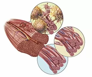
Muscle anatomy. Cutaway artwork showing the anatomy and internal structure of a muscle, from the macroscopic (left) to the microscopic (right) level

Background imageActin Collection: Movement of Listeria bacteria

Movement of Listeria bacteria
Movement of Listeria sp. bacteria. Immunofluorescence deconvolution micrograph showing the comet-like tails that help to propel Listeria sp. bacteria from cell to cell during an infection

Background imageActin Collection: Vaccinia virus infected cell

Vaccinia virus infected cell. Immunofluorescence deconvolution micrograph of a cell infected with vaccinia virus particles. Host and viral DNA (deoxyribonucleic acid) is blue

Background imageActin Collection: E. coli infection mechanism

E. coli infection mechanism. Immunofluorescence deconvolution micrograph of Escherichia coli bacteria (centre). Bacterial DNA (deoxyribonucleic acid) is blue

Background imageActin Collection: Fibroblast cells, fluorescent micrograph

Fibroblast cells, fluorescent micrograph
Fibroblast cells. Immunofluorescence deconvolution micrograph of fibroblast cells. The cell nuclei, which contain the cells genetic information, are blue


All products are expertly crafted, using premium materials, tailored to your specifications and promptly shipped


EDITORS COMMENTS

"Actin: The Dynamic Force Behind Cell Structure and Muscle Contraction" From HeLa cells to muscle fibers, actin plays a crucial role in various biological processes. This light micrograph C017 / 8299 showcases the intricate cell structure where actin filaments are prominently visible. In an immunofluorescent LM of fibroblast cell nuclei, actin's presence highlights its involvement in cellular movement and shape maintenance. Zooming into the intestinal microvilli through TEM reveals how actin filaments support their finger-like projections, aiding in nutrient absorption. Similarly, a confocal light micrograph of heart muscle unveils the organized arrangement and myosin filaments responsible for cardiac contractions. Stepping away from biology momentarily, we find ourselves at the 1973 24 Hours of Le Mans race. While seemingly unrelated, this event marked a breakthrough as researchers discovered similarities between car engines' pistons and muscle fiber structures - both relying on actin-myosin interactions for efficient performance. Returning to human anatomy, an artwork beautifully illustrates DTM (deep transverse massage) targeting deep back muscles affected by sprain, strain, or spasm. Actively engaging these muscles requires understanding their structure intimately. Delving deeper into muscle contraction mechanisms with labeled illustrations enlightens us about how actomyosin cross-bridges form during this process. Actins' ability to bind with myosins initiates muscular force generation necessary for any physical activity. Whether it's supporting cellular architecture or powering our movements during sports events like Le Mans or everyday activities like walking upstairs – "actin" remains an essential player behind the scenes.

© Copyright Media Storehouse, All Rights Reserved 2003 - 2025
Facebook     Pinterest     100% Payment Secure     Reviews IO