Fraud Blocker Skip to main content

Alveolus Collection

An alveolus, also known as a pulmonary alveolus, is a tiny air sac found in the human lungs

Background imageAlveolus Collection: Lactating breast tissue, light micrograph

Lactating breast tissue, light micrograph
Lactating breast tissue. Light micrograph of a section through healthy breast (mammary gland) tissue during lactation. Glandular tissue in the breast has proliferated to produce spaces (alveoli)

Background imageAlveolus Collection: Prostate, light micrograph

Prostate, light micrograph
Prostate. Light micrograph of a section through a prostate showing a concretion (red) of secretory materials in a prostatic alveolus (air space)

Background imageAlveolus Collection: Lung tissue, TEM

Lung tissue, TEM
Lung tissue, coloured transmission electron micrograph (TEM). A single alveolus is at center. At top right is a pulmonary capillary containing a red blood cell (red) and a platelet (yellow)

Background imageAlveolus Collection: Meadow brown butterfly, Maniola jurtina, wood lady or orange tip, Anthocharis cardamines

Meadow brown butterfly, Maniola jurtina, wood lady or orange tip, Anthocharis cardamines, and grizzled skipper
FLO4638853 Meadow brown butterfly, Maniola jurtina, wood lady or orange tip, Anthocharis cardamines, and grizzled skipper, Pyrgus alveolus

Background imageAlveolus Collection: Meadow brown butterfly, Maniola jurtina, wood lady or orange tip, Anthocharis cardamines

Meadow brown butterfly, Maniola jurtina, wood lady or orange tip, Anthocharis cardamines, and grizzled skipper, Pyrgus alveolus

Background imageAlveolus Collection: Diseased alveoli in the lung

Diseased alveoli in the lung. Cutaway computer artwork showing the interior of diseased alveoli (spheres) and bronchioles, (tubes) and blood vessels (red and purple) of the lungs

Background imageAlveolus Collection: Lung alveoli tree

Lung alveoli tree
Antique illustration of a Lung alveoli tree

Background imageAlveolus Collection: Illustration of respiratory system of bird showing lung, trachea, bronchus, pulmonary alveolus

Illustration of respiratory system of bird showing lung, trachea, bronchus, pulmonary alveolus, and syrinx

Background imageAlveolus Collection: Lung alveoli anatomy, artwork C016 / 7680

Lung alveoli anatomy, artwork C016 / 7680
Lung alveoli anatomy. Artwork showing the structure of the lung alveoli, where gas exchange occurs between the respiratory and circulatory systems

Background imageAlveolus Collection: Alveolus in human lung

Alveolus in human lung
Alveolus. False-colour scanning electron micrograph (SEM) of an alveolus (blue cavity), one of the microscopic air sacs of the lungs

Background imageAlveolus Collection: Lung alveoli

Lung alveoli. Light micrograph of a section through normal human lung tissue showing alveoli. These are tiny air sacs where most of the diffusion of gases to and from the blood occurs

Background imageAlveolus Collection: Bacterial lung infection

Bacterial lung infection, computer artwork. Rod-shaped bacteria (bacilli, green) on the surface of the alveoli of the lungs (purple)

Background imageAlveolus Collection: Pulmonary alveolus

Pulmonary alveolus
Antique illustration of a Pulmonary alveolus

Background imageAlveolus Collection: Illustration of human lung with small pulmonary alveolus surrounded by small particles causing

Illustration of human lung with small pulmonary alveolus surrounded by small particles causing inflammation to lining

Background imageAlveolus Collection: Illustration of development of idiopathic pulmonary fibrosis showing bronchus

Illustration of development of idiopathic pulmonary fibrosis showing bronchus and alveolus covered in small inorganic particles

Background imageAlveolus Collection: Illustration of bronchus and alveolus covered in inorganic particles, fibroblasts

Illustration of bronchus and alveolus covered in inorganic particles, fibroblasts, and growth factor obstructing the internal lining

Background imageAlveolus Collection: Illustration showing gas exchange in alveolus

Illustration showing gas exchange in alveolus

Background imageAlveolus Collection: Cross section biomedical illustration of alveolus

Cross section biomedical illustration of alveolus

Background imageAlveolus Collection: Cross-section of the alveoli branches

Cross-section of the alveoli branches

Background imageAlveolus Collection: Conceptual image of alveolus

Conceptual image of alveolus

Background imageAlveolus Collection: Conceptual image of alveoli

Conceptual image of alveoli

Background imageAlveolus Collection: Close-up of alveolus

Close-up of alveolus
The alveoli are the final branchings of the respiratory tree and act as the primary gas exchange units of the lung

Background imageAlveolus Collection: Conceptual image of human lungs

Conceptual image of human lungs

Background imageAlveolus Collection: Illustration of respiratory system

Illustration of respiratory system
Medicine: Human anatomy, respiratory system. Drawing

Background imageAlveolus Collection: Illustration of lobe of lung

Illustration of lobe of lung
Medicine: Human anatomy, lobe of the lung

Background imageAlveolus Collection: Alveoli

Alveoli
Medicine - Human Histology - Respiratory system - Lungs - Alveoli under microscope

Background imageAlveolus Collection: Lungs anatomy, artwork

Lungs anatomy, artwork
Lungs anatomy, computer artwork. At top left is the trachea (windpipe, white), which splits into two bronchi, one for each lung (blue)

Background imageAlveolus Collection: SEM of lung alveoli

SEM of lung alveoli
Lung alveoli. Coloured Scanning Electron Micro- graph (SEM) of lung tissue, showing alveoli and a pulmonary blood vessel. At centre is the lumen of a large blood vessel (yellow)

Background imageAlveolus Collection: Bronchioles

Bronchioles
Bronchiole network, composite image. The trachea (windpipe, not seen) branches into two bronchi, which enter the pair of lungs

Background imageAlveolus Collection: Lactating breast tissue, SEM

Lactating breast tissue, SEM

Background imageAlveolus Collection: Rat lung alveoli, SEM

Rat lung alveoli, SEM
Rat lung alveoli. Coloured scanning electron micrograph (SEM) of a section of a rat lung. Alveolar ducts (the larger passages) are surrounded by alveolar air sacs, which are clusters of alveoli

Background imageAlveolus Collection: Computer illustration showing alveoli

Computer illustration showing alveoli
Alveoli. Computer graphic illustration showing the alveoli (round light blue structures), the microscopic air sacs of the lungs

Background imageAlveolus Collection: Lung alveoli, SEM

Lung alveoli, SEM
Lung alveoli. Coloured scanning electron micrograph (SEM) of alveoli (air spaces) in the lungs. Blood vessels containing erythrocytes (red blood cells) are also seen

Background imageAlveolus Collection: F / col TEM of capillary in alveolar septum of lung

F / col TEM of capillary in alveolar septum of lung
False-colour transmission electron micrograph (TEM) of human lung tissue, showing an alveolar septum, the partition between the alveoli (air sacs) where gas exchange takes place

Background imageAlveolus Collection: LM of a cross section through alveoli in the lung

LM of a cross section through alveoli in the lung
Lung alveoli. Light micrograph of a cross section through lung tissue showing alveoli. Making up the frame (spongy appearance) are tiny air sacs called alveoli

Background imageAlveolus Collection: Coloured SEM of lung showing alveoli and bronchus

Coloured SEM of lung showing alveoli and bronchus
Lung alveoli & bronchus. Coloured Scanning Elec- tron Micrograph (SEM) of lung tissue, showing alveoli, a bronchus, and a pulmonary blood vessel

Background imageAlveolus Collection: Artwork of sputum in lungs with chronic bronchitis

Artwork of sputum in lungs with chronic bronchitis
Chronic bronchitis. Illustration of sputum collecting in the airways of a human lung affected by chronic bronchitis. The lobed structures are bunches of alveoli

Background imageAlveolus Collection: Artwork of asthmatic respiratory system on inhaler

Artwork of asthmatic respiratory system on inhaler
Asthma. Artwork of part of the respiratory system, seen on an aerosol inhaler (blue), showing the effects of asthma. The bronchi, covered in bands of cartilage (pink)

Background imageAlveolus Collection: Artwork of bronchodilator action in asthma

Artwork of bronchodilator action in asthma
Asthma treatment. Artwork showing the action of a bronchodilator drug breathed into the respiratory system from an aerosol inhaler to treat asthma

Background imageAlveolus Collection: Artwork of lung oedema in heart failure

Artwork of lung oedema in heart failure
Lung oedema in heart failure. Illustration showing leakage of fluid (oedema) into the lung due to heart failure. A magnified circle shows one of the lungs air sacs filled with fluid

Background imageAlveolus Collection: Lung fibrosis, CT scan

Lung fibrosis, CT scan
Lung fibrosis. Image 2 of 3. Coloured computed tomography (CT) scan through the chest of a patient with lung (pulmonary) fibrosis

Background imageAlveolus Collection: Asthma

Asthma. Computer artwork of allergens (blue and green) amongst alveoli (tiny air sacs, beige) in a lung. Allergens, such as dust and pollen, can trigger an asthma attack

Background imageAlveolus Collection: Artwork of the respiratory system of an asthmatic

Artwork of the respiratory system of an asthmatic
Asthma. Cut-away illustration of the respiratory system showing the effects of asthma. The trachea (windpipe) is at top and the alveoli, air sacs (white), are at bottom

Background imageAlveolus Collection: Lung blood vessel, SEM

Lung blood vessel, SEM
Lung blood vessel. Coloured scanning electron micrograph (SEM) of blood vessels around alveoli (air spaces, dark brown) in the lungs. Erythrocytes (red blood cells) are seen in the blood vessel

Background imageAlveolus Collection: Alveoli, artwork

Alveoli, artwork
Alveoli at the end of a bronchial tree, computer artwork

Background imageAlveolus Collection: Alveoli

Alveoli
3d medical illustration showing the alveoli and blood vessels in the human lung

Background imageAlveolus Collection: Human lungs, artwork

Human lungs, artwork
Human lungs, computer artwork

Background imageAlveolus Collection: Bronchial tree, artwork

Bronchial tree, artwork
Bronchial tree, computer artwork


All Professionally Made to Order for Quick Shipping

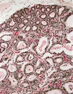
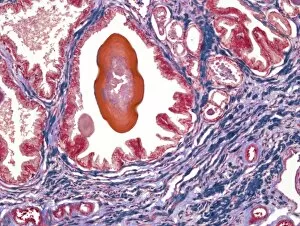
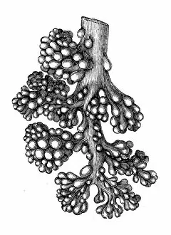
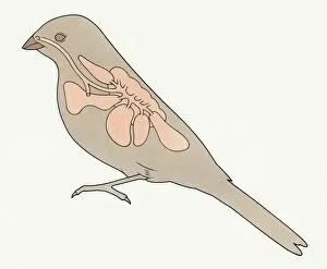
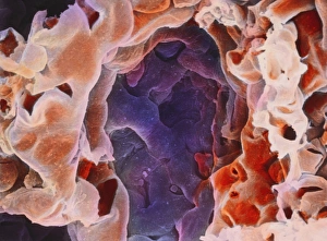
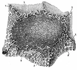
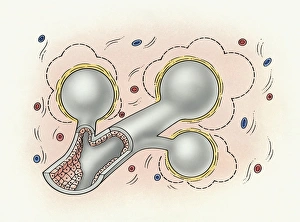
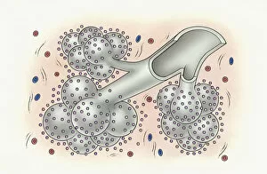
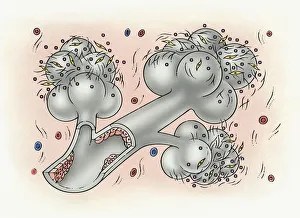
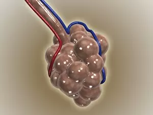
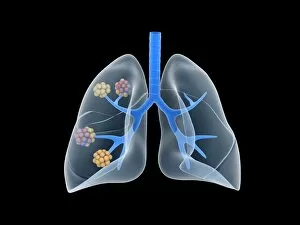
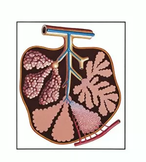
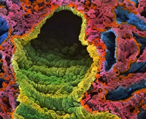
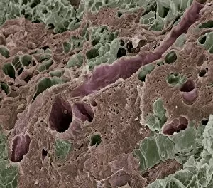
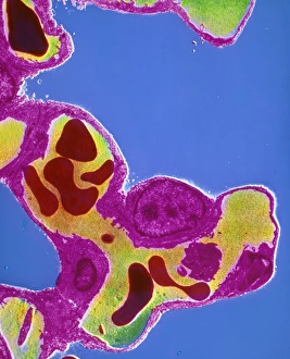
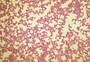
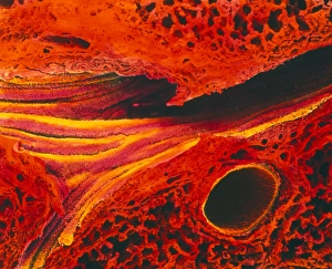
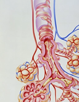
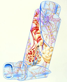
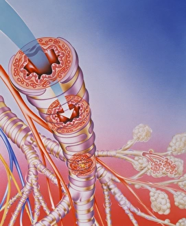
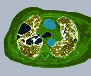
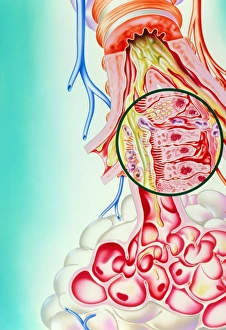
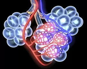
An alveolus, also known as a pulmonary alveolus, is a tiny air sac found in the human lungs. These microscopic structures play a crucial role in respiration by facilitating the exchange of oxygen and carbon dioxide between the bloodstream and the air. In healthy lungs, alveoli are lined with specialized cells that allow for efficient gas exchange. However, various factors can disrupt this delicate balance. For instance, lactating breast tissue can cause temporary changes to the structure of alveoli. Unfortunately, diseases such as bacterial lung infections can wreak havoc on these vital structures. When infected, alveoli become inflamed and may fill up with fluid or pus. This compromises their ability to function properly and leads to respiratory difficulties. The importance of maintaining healthy lung tissue cannot be overstated. In cases like idiopathic pulmonary fibrosis (IPF), small particles accumulate within bronchi and alveoli over time, causing inflammation and scarring. Fibroblasts and growth factors further obstruct the internal lining of these air sacs. Not only humans but birds also possess similar respiratory systems with intricate networks of bronchus and pulmonary alveoli called syrinxes. This enables them to produce complex vocalizations unique to each species. Understanding the structure and function of an alveolus is essential for comprehending respiratory health issues that arise when they are compromised or diseased. Through research and medical advancements, we strive towards preserving these tiny yet indispensable components of our lungs for optimal breathing capacity.